. 2024 Oct:76:103317.
doi: 10.1016/j.redox.2024.103317. Epub 2024 Aug 22.

AQP1 differentially orchestrates endothelial cell senescence

Affiliations

AQP1 differentially orchestrates endothelial cell senescence

Khatereh Shabanian et al. Redox Biol. 2024 Oct.

Abstract

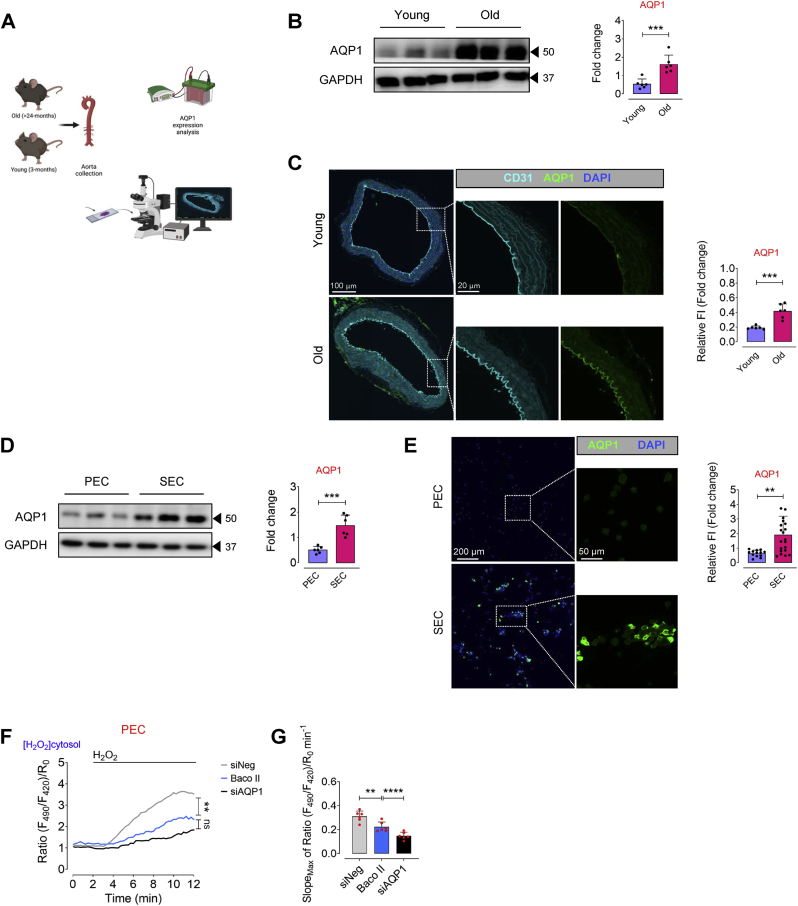
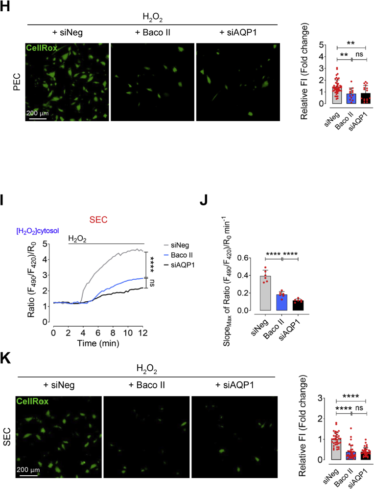
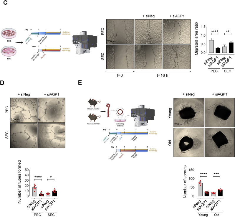
Accumulation of senescent endothelial cells (ECs) with age is a pivotal driver of cardiovascular diseases in aging. However, little is known about the mechanisms and signaling pathways that regulate EC senescence. In this report, we delineate a previously unrecognized role of aquaporin 1 (AQP1) in orchestrating extracellular hydrogen peroxide (H2O2)-induced cellular senescence in aortic ECs. Our findings underscore AQP1's differential impact on senescence hallmarks, including cell-cycle arrest, senescence-associated secretory phenotype (SASP), and DNA damage responses, intricately regulating angiogenesis. In proliferating ECs, AQP1 is crucial for maintaining angiogenic capacity, whereas disruption of AQP1 induces morphological and mitochondrial alterations, culminating in senescence and impaired angiogenesis. Conversely, Aqp1 knockdown or selective blockade of AQP1 in senescent ECs rescues the excess H2O2-induced cellular senescence phenotype and metabolic dysfunction, thereby ameliorating intrinsic angiogenic incompetence. Mechanistically, AQP1 facilitates H2O2 transmembrane transport, exacerbating oxidant-sensitive kinases CaMKII-AMPK. This process suppresses HDAC4 translocation, consequently de-repressing Mef2A-eNOS signaling in proliferating ECs. However, in senescent ECs, AQP1 overexpression is linked to preserved HDAC4-Mef2A complex and downregulation of eNOS signaling. Together, our studies identify AQP1 as a novel epigenetic regulator of HDAC4-Mef2A-dependent EC senescence and angiogenic potential, highlighting its potential as a therapeutic target for antagonizing age-related cardiovascular diseases.

Keywords: Aging; Angiogenesis; Aquaporin 1; Endothelial senescence; Epigenetic modification; Hydrogen peroxide.


Conflict of interest statement

Declaration of competing interest The authors declare the following financial interests/personal relationships which may be considered as potential competing interests: Soheil Saeedi reports was provided by University of Zurich. If there are other authors, they declare that they have no known competing financial interests or personal relationships that could have appeared to influence the work reported in this paper.

Figures

Image 1
Graphical abstract
Fig. 1
Fig. 1
Aging is associated with AQP1 upregulation in aortic endothelial cells. A, Schematic diagram of the experimental setting: Aortic ECs were collected from >24 (old) and 3 (young) -month-old C57BL/6 J male mice for AQP1 immunoblotting and immunostaining. B, Immunoblot analysis demonstrates a significant age-related increase in the expression of AQP1 in aortic ECs of mice (n = 6). C, AQP1 immunostaining demonstrates higher expression of AQP1 in the ascending aortic endothelial cells from old mice compared to those from young mice (n = 5–6). Bar chart reveals quantitative analysis of AQP1 fluorescence intensity normalized to CD31. D,E, Immunoblots and immunostaining represent a marked higher expression of AQP1 in senescent ECs (SEC; at p15-17) compared to proliferating ECs (PEC; at p4-5) (n = 6). Bar chart demonstrate quantitative analysis of AQP1 fluorescence intensity normalized to DAPI (n = 14–21). F, PECs were transduced with adenovirus 5 (AV5)-HyPer7.2 targeted to the cell cytosol for ratiometric fluorescence imaging to detect cytosolic H2O2 levels in the presence of exogeneous H2O2 (50 μM). The curves demonstrate significantly lower H2O2 responses in the cytosol of HyPer7.2-transduced PECs following treatment with either Baco II or siAQP1 as compared to that in siNeg-transfected PECs. G, Bar chart shows maximal HyPer7.2-ratio changes over time (slope) in the cytosol of PECs exposed to siNeg, Baco II, or siAQP1 (n = 6). H, CellRox green staining reveals a markedly lower generation of intracellular ROS, including H2O2, (as green fluorescence) in siNeg-transfected PECs as opposed to that in Baco II- or siAQP1-exposed PECs. Bar chart demonstrate quantitative analysis of CellRox fluorescence intensity normalized to DAPI (n = 13–47). I, SECs were transduced with AV5-HyPer7.2-Cyto for ratiometric fluorescence imaging to detect H2O2 in the cell cytosol in the presence of exogeneous H2O2 (50 μM). Both Baco II and siAQP1 significantly reduce H2O2 signals in the cytosol of HyPer7.2-expressing SECs compared to siNeg-PECs. J, Bar chart shows HyPer7.2 slope in the SECs exposed to siNeg, Baco II, or siAQP1 (n = 6). K, A significantly lower CellRox fluorescence intensity was seen in siNeg-SECs compared to Baco II- or siAQP1-exposed SECs. Bar chart demonstrate quantitative analysis of CellRox fluorescence intensity normalized to DAPI (n = 22–25). Scale bars, 20, 50, 100 and 200 μm. Error bars represent SD (B-E,G,H,J,K). Continuous data are presented as mean ± SD. Statistical analysis was performed with a two-tailed unpaired Student's t-test (B-E) and one-way ANOVA followed by Tukey's post hoc test (F,G,I,J). (**P < 0.01, ***P < 0.001, ****P < 0.0001, ns, not significant). Source data are provided as a Source Data file. (For interpretation of the references to colour in this figure legend, the reader is referred to the Web version of this article.)
Fig. 1
Fig. 1
Aging is associated with AQP1 upregulation in aortic endothelial cells. A, Schematic diagram of the experimental setting: Aortic ECs were collected from >24 (old) and 3 (young) -month-old C57BL/6 J male mice for AQP1 immunoblotting and immunostaining. B, Immunoblot analysis demonstrates a significant age-related increase in the expression of AQP1 in aortic ECs of mice (n = 6). C, AQP1 immunostaining demonstrates higher expression of AQP1 in the ascending aortic endothelial cells from old mice compared to those from young mice (n = 5–6). Bar chart reveals quantitative analysis of AQP1 fluorescence intensity normalized to CD31. D,E, Immunoblots and immunostaining represent a marked higher expression of AQP1 in senescent ECs (SEC; at p15-17) compared to proliferating ECs (PEC; at p4-5) (n = 6). Bar chart demonstrate quantitative analysis of AQP1 fluorescence intensity normalized to DAPI (n = 14–21). F, PECs were transduced with adenovirus 5 (AV5)-HyPer7.2 targeted to the cell cytosol for ratiometric fluorescence imaging to detect cytosolic H2O2 levels in the presence of exogeneous H2O2 (50 μM). The curves demonstrate significantly lower H2O2 responses in the cytosol of HyPer7.2-transduced PECs following treatment with either Baco II or siAQP1 as compared to that in siNeg-transfected PECs. G, Bar chart shows maximal HyPer7.2-ratio changes over time (slope) in the cytosol of PECs exposed to siNeg, Baco II, or siAQP1 (n = 6). H, CellRox green staining reveals a markedly lower generation of intracellular ROS, including H2O2, (as green fluorescence) in siNeg-transfected PECs as opposed to that in Baco II- or siAQP1-exposed PECs. Bar chart demonstrate quantitative analysis of CellRox fluorescence intensity normalized to DAPI (n = 13–47). I, SECs were transduced with AV5-HyPer7.2-Cyto for ratiometric fluorescence imaging to detect H2O2 in the cell cytosol in the presence of exogeneous H2O2 (50 μM). Both Baco II and siAQP1 significantly reduce H2O2 signals in the cytosol of HyPer7.2-expressing SECs compared to siNeg-PECs. J, Bar chart shows HyPer7.2 slope in the SECs exposed to siNeg, Baco II, or siAQP1 (n = 6). K, A significantly lower CellRox fluorescence intensity was seen in siNeg-SECs compared to Baco II- or siAQP1-exposed SECs. Bar chart demonstrate quantitative analysis of CellRox fluorescence intensity normalized to DAPI (n = 22–25). Scale bars, 20, 50, 100 and 200 μm. Error bars represent SD (B-E,G,H,J,K). Continuous data are presented as mean ± SD. Statistical analysis was performed with a two-tailed unpaired Student's t-test (B-E) and one-way ANOVA followed by Tukey's post hoc test (F,G,I,J). (**P < 0.01, ***P < 0.001, ****P < 0.0001, ns, not significant). Source data are provided as a Source Data file. (For interpretation of the references to colour in this figure legend, the reader is referred to the Web version of this article.)
Fig. 2
Fig. 2
AQP1 differentially regulates endothelial cell senescence. A, Schematic diagram of the experimental setting: PECs were treated with exogenous H2O2 (25 μM) for 24 h or Baco II (10 μM) for 72 h. Another subset of PECs was transfected with siNeg or siAQP1. At day 3, all cells were then subjected to senescence hallmarks profiling. B, Left, Upper panel, Representative images show significant increase in the numbers of SA-β-gal+ cells in the Baco II- and siAQP1-exposed groups, at the magnitude seen in H2O2–treated PECs, compared to siNeg-transfected PECs. Right, quantitative plots are shown for SA-β-gal+ cells (%) (n = 6). Left, Lower panel, γ-H2A.X immunostaining represent DDR in Baco II- and siAQP1-exposed PECs compared to siNeg-transfected PECs (n = 6). Quantitative γ-H2A.X fluorescence intensity normalized to DAPI are shown (n = 15–24). C, qPCR demonstrates that Baco II or siAQP1 treatment resulted in increased expression of CDK inhibitors p16INK4a, p19INK4d, and p21WAF1/Cip1 in PECs. D, qPCR shows that SASP components genes IL1α, IL-1β, and IL-6 are significantly upregulated in Baco II- and siAQP1-exposed PECs (n = 6). E, VCAM1 immunostaining shows its marked overexpression in Baco II- and siAQP1-exposed PECs compared to siNeg-transfected PECs (n = 6). F, Schematic diagram of the experimental setting: SECs were incubated with exogenous H2O2 (25 μM) for 24 h or Baco II (10 μM) for 72 h. Other SECs were transfected with siNeg or siAQP1. At day 3, all cells were then tested for senescence hallmarks. Left, Upper panel, Baco II or siAQP1 significantly decrease the numbers of SA-β-gal+ cells compared to that in siNeg-transfected SECs. Right, quantitative plots are shown for SA-β-gal+ cells (%) (n = 6). Left, Lower panel, γ-H2A.X immunostaining represent a markedly suppressed DDR in Baco II- and siAQP1-exposed SECs as opposed to that in siNeg-transfected SECs (n = 6). Bar chart demonstrates quantitative γ-H2A.X fluorescence intensity normalized to DAPI (n = 18–23). G, Baco II or siAQP1 restored cell cycle, represented by reduced p16INK4a, p19INK4d, and p21WAF1/Cip1 transcripts in SECs. H, A significant decrease in transcription of SASP components IL1α, IL-1β, and IL-6 was seen in Baco II- or siAQP1-exposed SECs compared to siNeg-transfected SECs (n = 6). I, VCAM1 immunostaining represents its marked downregulation in response to Baco II or siAQP1 in SECs as opposed to that in siNeg-transfected SECs (n = 6). Data from in vitro cellular experiments represent triplicated biologically independent experiments. Scale bar, 50 and 200 μm. Error bars represent SD (B-D, F–H). P values were calculated using one-way ANOVA followed by Tukey's post hoc test (B-D, F–H). (*P < 0.05, **P < 0.01, ***P < 0.001, ****P < 0.0001). Source data are provided as a Source Data file.
Fig. 2
Fig. 2
AQP1 differentially regulates endothelial cell senescence. A, Schematic diagram of the experimental setting: PECs were treated with exogenous H2O2 (25 μM) for 24 h or Baco II (10 μM) for 72 h. Another subset of PECs was transfected with siNeg or siAQP1. At day 3, all cells were then subjected to senescence hallmarks profiling. B, Left, Upper panel, Representative images show significant increase in the numbers of SA-β-gal+ cells in the Baco II- and siAQP1-exposed groups, at the magnitude seen in H2O2–treated PECs, compared to siNeg-transfected PECs. Right, quantitative plots are shown for SA-β-gal+ cells (%) (n = 6). Left, Lower panel, γ-H2A.X immunostaining represent DDR in Baco II- and siAQP1-exposed PECs compared to siNeg-transfected PECs (n = 6). Quantitative γ-H2A.X fluorescence intensity normalized to DAPI are shown (n = 15–24). C, qPCR demonstrates that Baco II or siAQP1 treatment resulted in increased expression of CDK inhibitors p16INK4a, p19INK4d, and p21WAF1/Cip1 in PECs. D, qPCR shows that SASP components genes IL1α, IL-1β, and IL-6 are significantly upregulated in Baco II- and siAQP1-exposed PECs (n = 6). E, VCAM1 immunostaining shows its marked overexpression in Baco II- and siAQP1-exposed PECs compared to siNeg-transfected PECs (n = 6). F, Schematic diagram of the experimental setting: SECs were incubated with exogenous H2O2 (25 μM) for 24 h or Baco II (10 μM) for 72 h. Other SECs were transfected with siNeg or siAQP1. At day 3, all cells were then tested for senescence hallmarks. Left, Upper panel, Baco II or siAQP1 significantly decrease the numbers of SA-β-gal+ cells compared to that in siNeg-transfected SECs. Right, quantitative plots are shown for SA-β-gal+ cells (%) (n = 6). Left, Lower panel, γ-H2A.X immunostaining represent a markedly suppressed DDR in Baco II- and siAQP1-exposed SECs as opposed to that in siNeg-transfected SECs (n = 6). Bar chart demonstrates quantitative γ-H2A.X fluorescence intensity normalized to DAPI (n = 18–23). G, Baco II or siAQP1 restored cell cycle, represented by reduced p16INK4a, p19INK4d, and p21WAF1/Cip1 transcripts in SECs. H, A significant decrease in transcription of SASP components IL1α, IL-1β, and IL-6 was seen in Baco II- or siAQP1-exposed SECs compared to siNeg-transfected SECs (n = 6). I, VCAM1 immunostaining represents its marked downregulation in response to Baco II or siAQP1 in SECs as opposed to that in siNeg-transfected SECs (n = 6). Data from in vitro cellular experiments represent triplicated biologically independent experiments. Scale bar, 50 and 200 μm. Error bars represent SD (B-D, F–H). P values were calculated using one-way ANOVA followed by Tukey's post hoc test (B-D, F–H). (*P < 0.05, **P < 0.01, ***P < 0.001, ****P < 0.0001). Source data are provided as a Source Data file.
Fig. 3
Fig. 3
AQP1 differentially orchestrates endothelial energy homeostasis and angiogenesis. Mitochondrial respiratory rates in PECs (A) and SECs (B) exposed to siNeg, H2O2 alone (25 μM), Baco II, or siAQP1 was measured using Seahorse flux analyzer by Cell Mito Stress kit (n = 6). Bar charts reveal significant reduction in oxygen consumption rate (OCR) represented by decreased maximal, basal, and spare reserve, and ATP biosynthesis in Baco II- or siAQP1-exposed PECs compared to PECs transfected with siNeg. Moreover, H2O2-exposed PECs exhibit significantly lower OCR measures compared to siNeg-transfected counterparts. Conversely, Baco II and siAQP1 restored mitochondrial respiration and markedly increased the OCR parameters in SECs as opposed to those in either siNeg-transfected or H2O2-treated SECs (n = 6). C, Phase-contrast micrographs depict that siAQP1 significantly reduces cell migration in PECs, shown as larger areas of uncovered surface, to the comparable level seen in siNeg-transfected SECs. While, SECs transfected with siAQP1 exhibited a marked increase in cell migration compared to siNeg-SECs. Right, Bar chart represents the ratio of cell migrated area (n = 6). D, Phase-contrast images represent the 2-D matrigel tube formation of PECs (Upper) and SECs (Lower) transfected with siNeg or siAQP1. Right, quantitative plot shows a significantly lower number of tubes formed by siAQP1-transfected PECs, while a markedly higher number of tubes was observed in siAQP1-SEC group compared to siNeg-transfected cells (n = 6). E, Phase-contrast micrographs of the aortic rings from old and young mice show that siAQP1 transfection of young aortas significantly decreases endothelial sprouting, whereas markedly increases angiogenic capacity in old aortas compared to siNeg-transfected aortas. Bottom, quantitative plot is shown for the number of aortic endothelial sprouts (n = 6). Scale bars, 200 μm. Data were determined in 6 micrographs from 3 different plates and represent triplicated biologically independent experiments. Error bars represent SD (A-E). P values were calculated using one-way (A,B) and two-way (C-E) ANOVA followed by Tukey's post hoc test. (*P < 0.05, **P < 0.01, ***P < 0.001, ****P < 0.0001, ns, not significant). Source data are provided as a Source Data file.
Fig. 3
Fig. 3
AQP1 differentially orchestrates endothelial energy homeostasis and angiogenesis. Mitochondrial respiratory rates in PECs (A) and SECs (B) exposed to siNeg, H2O2 alone (25 μM), Baco II, or siAQP1 was measured using Seahorse flux analyzer by Cell Mito Stress kit (n = 6). Bar charts reveal significant reduction in oxygen consumption rate (OCR) represented by decreased maximal, basal, and spare reserve, and ATP biosynthesis in Baco II- or siAQP1-exposed PECs compared to PECs transfected with siNeg. Moreover, H2O2-exposed PECs exhibit significantly lower OCR measures compared to siNeg-transfected counterparts. Conversely, Baco II and siAQP1 restored mitochondrial respiration and markedly increased the OCR parameters in SECs as opposed to those in either siNeg-transfected or H2O2-treated SECs (n = 6). C, Phase-contrast micrographs depict that siAQP1 significantly reduces cell migration in PECs, shown as larger areas of uncovered surface, to the comparable level seen in siNeg-transfected SECs. While, SECs transfected with siAQP1 exhibited a marked increase in cell migration compared to siNeg-SECs. Right, Bar chart represents the ratio of cell migrated area (n = 6). D, Phase-contrast images represent the 2-D matrigel tube formation of PECs (Upper) and SECs (Lower) transfected with siNeg or siAQP1. Right, quantitative plot shows a significantly lower number of tubes formed by siAQP1-transfected PECs, while a markedly higher number of tubes was observed in siAQP1-SEC group compared to siNeg-transfected cells (n = 6). E, Phase-contrast micrographs of the aortic rings from old and young mice show that siAQP1 transfection of young aortas significantly decreases endothelial sprouting, whereas markedly increases angiogenic capacity in old aortas compared to siNeg-transfected aortas. Bottom, quantitative plot is shown for the number of aortic endothelial sprouts (n = 6). Scale bars, 200 μm. Data were determined in 6 micrographs from 3 different plates and represent triplicated biologically independent experiments. Error bars represent SD (A-E). P values were calculated using one-way (A,B) and two-way (C-E) ANOVA followed by Tukey's post hoc test. (*P < 0.05, **P < 0.01, ***P < 0.001, ****P < 0.0001, ns, not significant). Source data are provided as a Source Data file.
Fig. 4
Fig. 4
Differential AQP1-mediated endothelial senescence is elicited by HDAC4-Mef2A pathway. A-F, Immunoblot analysis reveals the differential role of AQP1 in the regulation of CaMKII-AMPK-mediated phosphorylation of HDAC4 at Ser632 and the subsequent Mef2A-eNOS phosphorylation in PECs versus SECs (n = 6). Data revealed that H2O2 (25 μM) markedly increases CaMKII-mediated phosphorylation of AMPK, leading to HDAC4S632 phosphorylation and an increased expression of Mef2A. This sequence reinforces eNOS phosphorylation at Ser1177 in PECs. However, silencing (siAQP1) or inhibiting (Baco II) AQP1 blocked the permeability to H2O2, reducing CAMKII-AMPK-mediated phosphorylation of HDAC4S632, and subsequently downregulating eNOSS1177 phosphorylation in these cells. In contrast, in SECs exposed to siNeg or exogenous H2O2 (25 μM), there was a marked reduction in CaMKII-AMPK phosphorylation, thereby leading to a decreased in Mef2A expression and further eNOSS1177 phosphorylation. These CaMKII-regulated epigenetic alterations and the subsequent eNOS dephosphorylation were reversed by either siAQP1 or Baco II in SECs. G, Immunoblots (left) and quantitative plots (right) reveal that siAQP1 stimulates nuclear localization of HDAC4, represented as increased expression in the nucleus, in PECs (top), while it facilitates nuclear export, represented as a decrease in HDAC4 expression in the cytosol, in SECs (bottom) (n = 6). H,p-HDAC4 immunostaining shows that siAQP1 markedly decreases HDAC4 phosphorylation, facilitating its nuclear localization in PECs. However, siAQP1 transfection facilitates HDAC4 translocation towards the cell cytosol, shown as higher pHDAC4 fluorescence, in SECs (n = 6). Right, quantitative p-HDAC4S632 fluorescence intensity normalized to DAPI are shown (n = 19–28). I, The mechanism underlying the differential contribution of AQP1 to endothelial cell senescence and angiogenic capacity. AQP1 differentially orchestrates H2O2-mediated EC senescence. (Left) In PECs, AQP1 is crucial for H2O2-regulated CaMKII-AMPK stimulation and the subsequent HDAC4 phosphorylation (nuclear export), facilitating Mef2A expression and eNOSS1177 phosphorylation. Aqp1 silencing or pharmacological inhibition of AQP1 promotes cellular senescence and impairs angiogenesis in these cells. (Right) In SECs, higher AQP1 abundance contributes to oxidative stress that suppresses CaMKII-AMPK-mediated HDAC4 translocation, subsequently leading to reduced Mef2A expression and eNOS activation. Conversely, AQP1 deficiency reduces intracellular ROS accumulation and thereby both reverses cellular senescence and restores angiogenic capacity in these cells. Scale bars, 50 μm. Data were determined in 6 micrographs from 3 different plates and represent triplicated biologically independent experiments. Error bars represent SD (B-G). Continuous data are presented as mean ± SD. Statistical analysis was performed with a two-tailed unpaired Student's t-test (H) and one-way ANOVA followed by Tukey's post hoc test (B–F). (*P < 0.05, **P < 0.01, ***P < 0.001, ns, not significant). Source data are provided as a Source Data file.
Fig. 4
Fig. 4
Differential AQP1-mediated endothelial senescence is elicited by HDAC4-Mef2A pathway. A-F, Immunoblot analysis reveals the differential role of AQP1 in the regulation of CaMKII-AMPK-mediated phosphorylation of HDAC4 at Ser632 and the subsequent Mef2A-eNOS phosphorylation in PECs versus SECs (n = 6). Data revealed that H2O2 (25 μM) markedly increases CaMKII-mediated phosphorylation of AMPK, leading to HDAC4S632 phosphorylation and an increased expression of Mef2A. This sequence reinforces eNOS phosphorylation at Ser1177 in PECs. However, silencing (siAQP1) or inhibiting (Baco II) AQP1 blocked the permeability to H2O2, reducing CAMKII-AMPK-mediated phosphorylation of HDAC4S632, and subsequently downregulating eNOSS1177 phosphorylation in these cells. In contrast, in SECs exposed to siNeg or exogenous H2O2 (25 μM), there was a marked reduction in CaMKII-AMPK phosphorylation, thereby leading to a decreased in Mef2A expression and further eNOSS1177 phosphorylation. These CaMKII-regulated epigenetic alterations and the subsequent eNOS dephosphorylation were reversed by either siAQP1 or Baco II in SECs. G, Immunoblots (left) and quantitative plots (right) reveal that siAQP1 stimulates nuclear localization of HDAC4, represented as increased expression in the nucleus, in PECs (top), while it facilitates nuclear export, represented as a decrease in HDAC4 expression in the cytosol, in SECs (bottom) (n = 6). H,p-HDAC4 immunostaining shows that siAQP1 markedly decreases HDAC4 phosphorylation, facilitating its nuclear localization in PECs. However, siAQP1 transfection facilitates HDAC4 translocation towards the cell cytosol, shown as higher pHDAC4 fluorescence, in SECs (n = 6). Right, quantitative p-HDAC4S632 fluorescence intensity normalized to DAPI are shown (n = 19–28). I, The mechanism underlying the differential contribution of AQP1 to endothelial cell senescence and angiogenic capacity. AQP1 differentially orchestrates H2O2-mediated EC senescence. (Left) In PECs, AQP1 is crucial for H2O2-regulated CaMKII-AMPK stimulation and the subsequent HDAC4 phosphorylation (nuclear export), facilitating Mef2A expression and eNOSS1177 phosphorylation. Aqp1 silencing or pharmacological inhibition of AQP1 promotes cellular senescence and impairs angiogenesis in these cells. (Right) In SECs, higher AQP1 abundance contributes to oxidative stress that suppresses CaMKII-AMPK-mediated HDAC4 translocation, subsequently leading to reduced Mef2A expression and eNOS activation. Conversely, AQP1 deficiency reduces intracellular ROS accumulation and thereby both reverses cellular senescence and restores angiogenic capacity in these cells. Scale bars, 50 μm. Data were determined in 6 micrographs from 3 different plates and represent triplicated biologically independent experiments. Error bars represent SD (B-G). Continuous data are presented as mean ± SD. Statistical analysis was performed with a two-tailed unpaired Student's t-test (H) and one-way ANOVA followed by Tukey's post hoc test (B–F). (*P < 0.05, **P < 0.01, ***P < 0.001, ns, not significant). Source data are provided as a Source Data file.

References

    1. Bloom S.I., et al. Mechanisms and consequences of endothelial cell senescence. Nat. Rev. Cardiol. 2023;20:38–51. - PMC - PubMed
    1. Allemann M.S., et al. Targeting the redox system for cardiovascular regeneration in aging. Aging Cell. 2023;22 - PMC - PubMed
    1. Gasek N.S., et al. Strategies for targeting senescent cells in human disease. Nat. Aging. 2021;1:870–879. - PMC - PubMed
    1. Han Y., Kim S.Y. Endothelial senescence in vascular diseases: current understanding and future opportunities in senotherapeutics. Exp. Mol. Med. 2023;55:1–12. - PMC - PubMed
    1. Yrrell D.J., Goldstein D.R. Ageing and atherosclerosis: vascular intrinsic and extrinsic factors and potential role of IL-6. Nat. Rev. Cardiol. 2021;18:58–68. - PMC - PubMed

Publication types

LinkOut - more resources