. 2024 Oct:76:103322.
doi: 10.1016/j.redox.2024.103322. Epub 2024 Aug 20.

GHSR deficiency exacerbates Parkinson's disease pathology by impairing autophagy

Affiliations

GHSR deficiency exacerbates Parkinson's disease pathology by impairing autophagy

Xue Xiao et al. Redox Biol. 2024 Oct.

Abstract

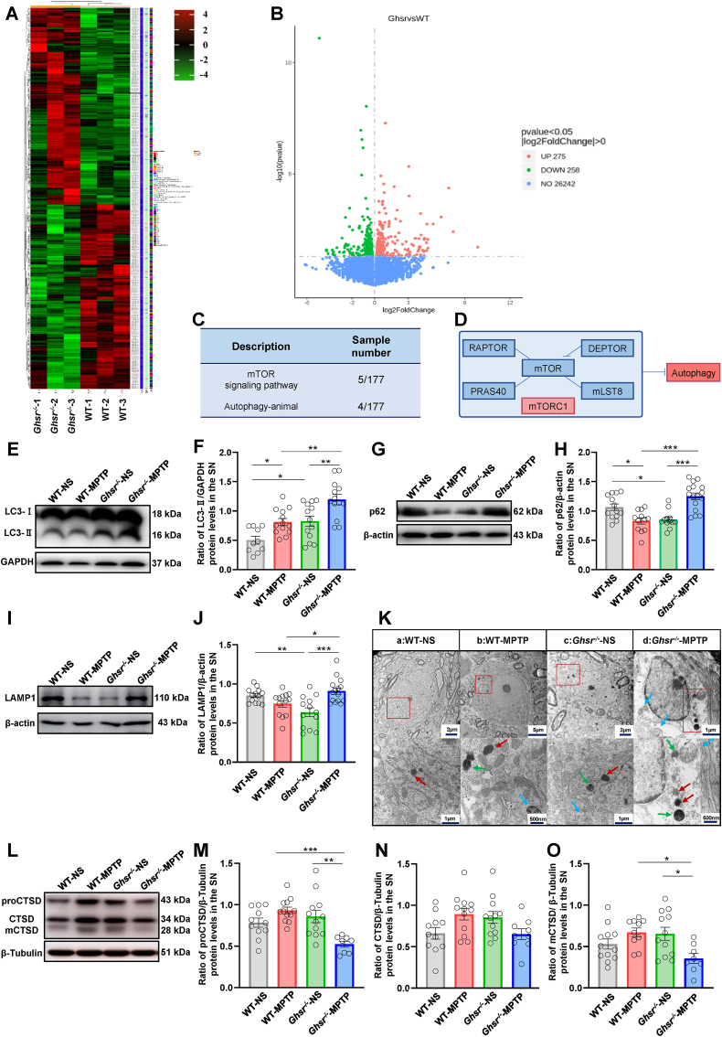
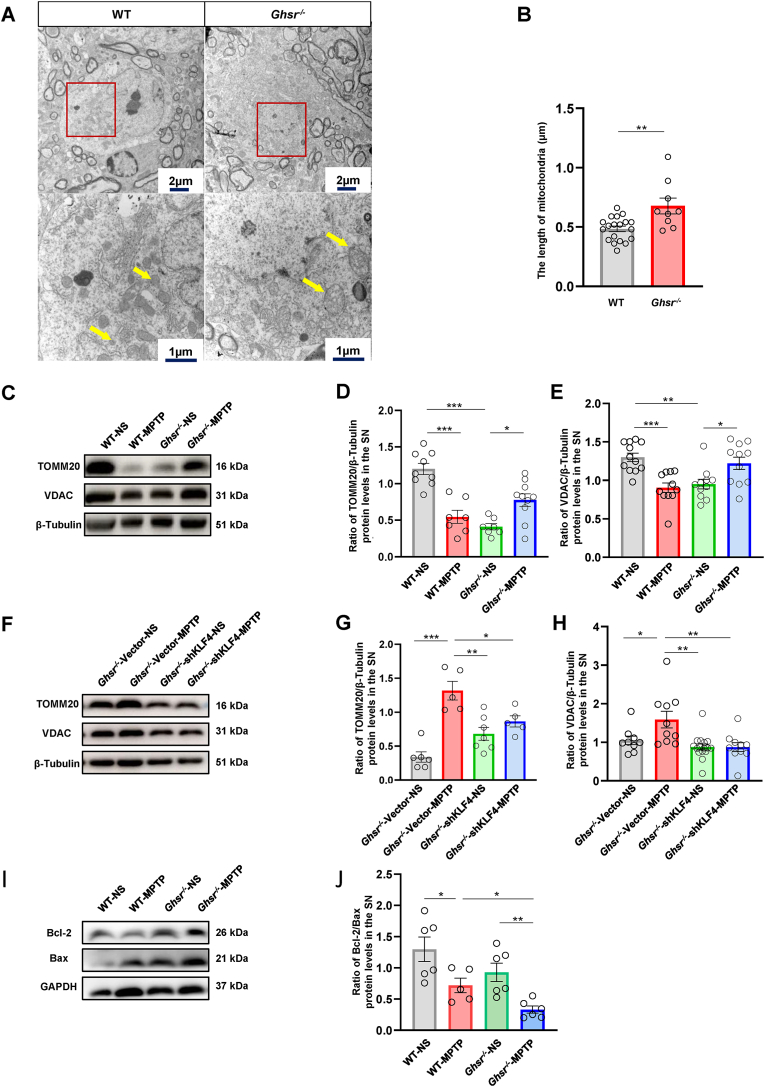
In Parkinson's disease (PD), exogenous ghrelin protects dopaminergic neurons through its receptor, growth hormone secretagogue receptor (GHSR). However, in contrast to the strikingly low levels of ghrelin, GHSR is highly expressed in the substantia nigra (SN). What role does GHSR play in dopaminergic neurons is unknown. In this study, using GHSR knockout mice (Ghsr-/- mice) and 1-methyl-4-phenyl-1,2,3,6-tetrahydropyridine (MPTP)-induced PD model, we found that GHSR deletion aggravated dopaminergic neurons degeneration, and the expression and activity of GHSR were significantly reduced in PD. Furthermore, we explored the potential mechanism that GHSR deficiency aggregated PD-related neurodegeneration. We showed that DEPTOR, a subunit of mTORC1, was overexpressed in Ghsr-/- mice, positively regulating autophagy and enhancing autophagy initiation. The expression of lysosomal markers was abnormal, implying lysosomal dysfunction. As a result, the damaged mitochondria could not be effectively eliminated, which ultimately exacerbated the injury of nigral dopaminergic neurons. In particular, we demonstrated that DEPTOR could be transcriptionally regulated by KLF4. Specific knockdown of KLF4 in dopaminergic neurons effectively alleviated neurodegeneration in Ghsr-/- mice. In summary, our results suggested that endogenous GHSR deletion-compromised autophagy by impairing lysosomal function, is a key contributor to PD, which provided ideas for therapeutic approaches involving the manipulation of GHSR.

Keywords: Autophagy; DEPTOR; GHSR; KLF4; Parkinson's disease.


Conflict of interest statement

Declaration of competing interest The authors have no other relevant affiliations or financial involvement with any organization or entity with a financial interest in or financial conflict with the subject matter or materials discussed in the manuscript apart from those disclosed.

Figures

Image 1
Graphical abstract
Fig. 1
Fig. 1
GHSR knockout aggravated dopaminergic neuron degeneration in PD mice A: Representative images showing TH-positive neurons in the SN in the different groups following immunofluorescence staining. Scale bar = 100 μm. B: Group data showing the number of TH-positive neurons in the different groups. C–E: Group data showing the levels of striatal DA, DOPAC and HVA in the different groups. F–H: Behavioral changes in the different groups were detected by the pole test and rotarod test. I: Representative images showing microglia in the SN in the different groups following immunofluorescent staining. Scale bar = 100 μm. J: Group data showing the number of microglia in the different groups. K–M: Western blotting was used to measure TH and SOD1 protein levels in the SN. Each bar represents the mean ± S.E.M. (one-way ANOVA; *P<0.05, **P<0.01, ***P<0.001).
Fig. 2
Fig. 2
The expression and activity of GHSR were decreased in PD A: The effect of MPP+ on GHSR protein expression in N2a cells. B: The effect of MPTP on GHSR protein expression in the SN of mice. C: The standard curve of IP. D: Schematic diagram of HTRF technology detecting the intracellular IP content. E: The effect of MPP+ on the intracellular IP content. Each bar represents the mean ± S.E.M. (one-way ANOVA, Student's t-test; *P<0.05, **P<0.01).
Fig. 3
Fig. 3
GHSR knockout enhanced autophagy initiation and blocked autophagic flux in PD mice A–B: Analysis of the transcriptomic results. C–D: Signaling pathways identified according to the transcriptomic results. E–J: Western blotting was used to measure the protein levels of LC3, p62, and LAMP1 in the SN of WT and Ghsr−/− mice. K: The morphological characteristics of the autophagosomes, lysosomes and autophagosomes in dopaminergic neurons in the SN were observed via transmission electron microscopy. The blue arrows represent autophagosomes, the red arrows represent lysosomes, and the green arrows represent autolysosomes. L–O: Western blotting was used to measure the protein level of CTSD in the SN of WT and Ghsr−/− mice. Each bar represents the mean ± S.E.M. (one-way ANOVA; *P<0.05, **P<0.01, ***P<0.001). (For interpretation of the references to color in this figure legend, the reader is referred to the Web version of this article.)
Fig. 4
Fig. 4
GHSR knockout-induced autophagy dysregulation was associated with the mTORC1 subunit DEPTOR A: Constituent subunits of mTORC1. B: The mRNA expression levels of the five subunits of mTORC1 in WT mice and Ghsr−/− mice were verified via RT-PCR. C–D: Western blotting was used to measure the protein levels of DEPTOR in the SN. E–L: The protein expression levels of RAPTOR, mLST8, PRAS40, and mTOR in WT mice and Ghsr−/− mice were verified by western blotting. Each bar represents the mean ± S.E.M. (one-way ANOVA, Student's t-test; *P<0.05, **P<0.01, ***P<0.001).
Fig. 5
Fig. 5
The transcription factor KLF4 could bind to the promoter of DEPTOR A: The transcription factor upstream of DEPTOR was predicted to be KLF4 by bioinformatics methods. B–C: Dual-luciferase reporter gene experiments confirmed that KLF4 and DEPTOR could bind to each other. D–H: The Biacore T200 instrument was used to analyze the interaction between DEPTOR and KLF4. KD was 5.083*10^ (−8), and the binding site sequence was 5′-CCTTGGTCTA-3'. I: The dual-luciferase reporter gene experiment confirmed that KLF4-DEPTOR binding ability was decreased after the promoter of DEPTOR was mutated. Each bar represents the mean ± S.E.M. (one-way ANOVA; ***P<0.001).
Fig. 6
Fig. 6
Knocking down KLF4 in dopaminergic neurons prevented MPTP-induced neurodegeneration in Ghsr−/− mice A: The protein expression of KLF4 increased after GHSR gene knockout. B: Experimental design for stereotactic injection of AAV-shKLF4 into the SN of Ghsr−/− mice. C: Western blotting was used to measure the protein level of KLF4 after AAV-shKLF4 injection. D–F: The movement of Ghsr−/− mice after stereotactic injection of AAV-shKLF4 was assessed via the pole test and rotarod test. G–J: Western blotting was used to measure the expressions of TH and LC3 in the SN of Ghsr−/− mice after stereotactic injection of AAVshKLF4. K–N: Western blotting was used to measure the expression of p62 and DEPTOR in the SN of Ghsr−/− mice after stereotactic injection of AAV-shKLF4. O–R: Western blotting was used to measure the expression of CTSD in the SN of Ghsr−/− mice after stereotactic injection of AAV-shKLF4. Each bar represents the mean ± S.E.M. (one-way ANOVA; *P<0.05, **P<0.01, ***P<0.001).
Fig. 7
Fig. 7
GHSR knockout-induced autophagy dysregulation inhibited mitochondrial clearance in PD mice A–B: Mitochondria in dopaminergic neurons in the SN of WT and Ghsr−/− mice were observed via transmission electron microscopy. C–H: Western blotting was used to measure the expression of TOMM20 and VDAC in the SN of WT and Ghsr−/− mice. I–J: Western blotting was used to measure the Bcl-2/Bax ratio in the SN. Each bar represents the mean ± S.E.M. (one-way ANOVA; *P<0.05, **P<0.01, ***P<0.001).

References

    1. Goldman S.M. Environmental toxins and Parkinson's disease. Annu. Rev. Pharmacol. Toxicol. 2014;54:141–164. - PubMed
    1. Samii A., Nutt J.G., Ransom B.R. Parkinson's disease. Lancet. 2004;363(9423):1783–1793. - PubMed
    1. Ascherio A., Schwarzschild M.A. The epidemiology of Parkinson's disease: risk factors and prevention. Lancet Neurol. 2016;15(12):1257–1272. - PubMed
    1. Song N., et al. Assessments of plasma ghrelin levels in the early stages of Parkinson's disease. Mov. Disord. 2017;32(10):1487–1491. - PubMed
    1. Yu J., et al. Ghrelin protects MES23.5 cells against rotenone via inhibiting mitochondrial dysfunction and apoptosis. Neuropeptides. 2016;56:69–74. - PubMed

Publication types