. 2024 Aug 24;15(1):7303.
doi: 10.1038/s41467-024-51566-5.

PHF6 cooperates with SWI/SNF complexes to facilitate transcriptional progression

Affiliations

PHF6 cooperates with SWI/SNF complexes to facilitate transcriptional progression

Priya Mittal et al. Nat Commun. .

Abstract

Genes encoding subunits of SWI/SNF (BAF) chromatin remodeling complexes are mutated in nearly 25% of cancers. To gain insight into the mechanisms by which SWI/SNF mutations drive cancer, we contributed ten rhabdoid tumor (RT) cell lines mutant for SWI/SNF subunit SMARCB1 to a genome-scale CRISPR-Cas9 depletion screen performed across 896 cell lines. We identify PHF6 as specifically essential for RT cell survival and demonstrate that dependency on Phf6 extends to Smarcb1-deficient cancers in vivo. As mutations in either SWI/SNF or PHF6 can cause the neurodevelopmental disorder Coffin-Siris syndrome, our findings of a dependency suggest a previously unrecognized functional link. We demonstrate that PHF6 co-localizes with SWI/SNF complexes at promoters, where it is essential for maintenance of an active chromatin state. We show that in the absence of SMARCB1, PHF6 loss disrupts the recruitment and stability of residual SWI/SNF complex members, collectively resulting in the loss of active chromatin at promoters and stalling of RNA Polymerase II progression. Our work establishes a mechanistic basis for the shared syndromic features of SWI/SNF and PHF6 mutations in CSS and the basis for selective dependency on PHF6 in SMARCB1-mutant cancers.


Conflict of interest statement

The authors declare no competing interests.

Figures
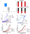
Fig. 1
Fig. 1. Genome-scale CRISPR-Cas9 screen identifies PHF6 as a dependency in rhabdoid tumor cell lines.
a Two-class comparison of 10 biologically independent RT cell lines (blue) versus n = 886 biologically independent other cell lines (gray) plotted with CERES score for dependency on PHF6 on the x-axis. Negative CERES score demonstrates increased dependency. Statistical analysis was performed using a Benjamini–Hochberg corrected two-tailed Student’s t-test; P = 1.12 × 10 − 8. b Indel frequency assay. PHF6 was targeted with a guide RNA-Cas9 RNP in G401 RT cells and control Pfeiffer cells, and genomic DNA was harvested and sequenced after 7, 14, and 21 days. All indels were binned into in-frame (red) or out-of-frame (black). A non-targeting guide was used as a control. Data are represented from mean of three biological replicates. c, d Effects of PHF6 knockdown on proliferation of SMARCB1-deficient RT cell lines or (d) SMARCB1-expressing ES2 and MCF7 control cell lines. Data are represented from mean of n = 16 technical replicates per cell line per condition from one independent experiment. e Effects of exogenous re-expression of PHF6 following knockdown of PHF6 in G401 and TTC549 cells. Data are represented from mean of n = 8 technical replicates per cell line per condition from one independent experiment. Source data are provided as a Source Data file.
Fig. 2
Fig. 2. PHF6 is required for maintenance of H3K14 and H3K27 acetylation.
a Volcano plot displaying histone post-translational modification changes as determined by mass spectrometry of histones extracted from G401 shPHF6 versus shCTRL cells. Two-tailed homoscedastic Student’s t-test was performed. Data are represented from mean of three biological replicates. b Western blots of histone modifications in histone extracts prepared from RT cells 72 hr post selection with shCTRL or shPHF6 knockdown (n = 3 independent biological replicates). c Western blots of histone modifications in G401 cells re-expressing inducible exogenous PHF6 (72 h) after knockdown of endogenous PHF6 (n = 2 independent biological replicates). d Metagene plots of H3K14ac, H3K27ac centered at the TSS ± 2 kb in shCTRL and shPHF6 G401 cells (n = 3 independent biological replicates). e Re-expression of doxycycline-inducible exogenous PHF6 (72 h) in DND41 cells that naturally lack PHF6 expression (n = 2 independent biological replicates). Source data are provided as a Source Data file.
Fig. 3
Fig. 3. PHF6 regulates histone acetyltransferase p300 and maintains chromatin accessibility.
a Distribution of PHF6 occupancy across chromatin states in G401 RT cells (n = 2 independent biological replicates). b Co-immunoprecipitation of PHF6 with p300 from nuclear extracts of G401 RT cells (n = 3 independent biological replicates). c Western blot analysis p300 protein levels in nuclear extracts from G401 RT cells after knockdown of PHF6 (n = 3 independent biological replicates). d Density heatmap of chromatin occupancy of PHF6, p300, H3K14ac, and H3K27ac in shCTRL and shPHF6 conditions in G401 (ordered by PHF6 occupancy). e Density heatmap of significant losses (log2FC < 0, FDR < 0.05) and gains (log2FC > 0, FDR < 0.05) in accessibility and chromatin occupancy changes of H3K14ac and H3K27ac upon loss of PHF6 ranked based on PHF6 binding are shown in G401 cells (n = 3 independent biological replicates). Promoter regions are designated by the presence of H3K4me3. f Example loci reflecting loss of H3K14ac, H3K27ac, p300, and resultant accessibility at PHF6-bound promoters. Source data are provided as a Source Data file.
Fig. 4
Fig. 4. PHF6 is required for promoter-proximal pause release of RNA polymerase II.
a PHF6 co-immunoprecipitation of Pol II and its phosphorylated forms in G401 RT cells (n = 2 independent biological replicates) b Western blot analysis of the levels of Pol II Ser5P (promoter proximal) and Pol II Ser2P (elongating) forms in shCTRL and shPHF6 G401 cells (n = 3 independent biological replicates). Metagene plots of (c) Pol II Ser5P (promoter proximal) and d Pol II Ser2P (elongating) in shCTRL (black) and shPHF6 (red) conditions (n = 2 independent biological replicates) at PHF6-bound genes (n = 8728). e Metagene nascent transcriptional profiles (Bru-seq read density) of PHF6 bound genes stratified based on gene-length (10–60 kb) at the 5’ (TSS) of protein-coding genes in shCTRL and shPHF6 conditions in G401 cells (n = 3 independent biological replicates). f Example locus depicting Pol II Ser5P, Pol II Ser2P, and nascent transcription upon loss of PHF6 at PHF6-bound SMAD9. g Gene length distributions (log10 transformed) for all hg19 genes (blue) and 345 significantly downregulated PHF6 target genes defined in RT cell lines (TTC709, TTC549, G401) in magenta. h Scatter plot displaying correlation between differentially expressed genes from Bru-seq and mRNA-seq upon PHF6 knockdown in G401 cells. i Representative locus depicting Pol II Ser5P, Pol II Ser2P, nascent transcript, and mRNA upon loss of PHF6 at PHF6-bound Jun locus. j Gene ontology analysis by Metascape of significantly downregulated and upregulated genes upon PHF6 knockdown (adjusted p < 0.05 and log2FC < 0 or log2FC > 0) in G401 cells. Source data are provided as a Source Data file.
Fig. 5
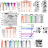
Fig. 5. PHF6 facilitates SWI/SNF function.
a Density heatmap of chromatin occupancy (ordered by PHF6) for PHF6, SMARCA4, PBRM1, BRD9, and H3K14ac in G401 cells (n = 2 independent biological replicates). b Chromatin occupancy peaks of PHF6, BRD9, PBRM1, and SMARCA4 overlapping with chromatin states defined based on ChromHMM. c PHF6 immunoprecipitation of SWI/SNF subunits from G401 nuclear extracts (n = 3 independent biological replicates) d Western blot analysis of SWI/SNF complex members from shCTRL and shPHF6 G401 nuclear lysates (n = 3 independent biological replicates). e Density sedimentation assays using 10–30% glycerol gradients performed on G401 lysates in shCTRL and shPHF6 conditions (n = 2 independent biological replicates). f Density heatmap in shCTRL and shPHF6 conditions (rank ordered by PHF6) at sites where H3K14ac is lost upon knockdown of PHF6 in G401 cells (n = 2 independent biological replicates). g Overlap of sites where BRD9, PBRM1, and SMARCA4 peaks are lost upon PHF6 knockdown in G401 with chromatin states defined based on ChromHMM. h Example locus reflecting reduced occupancy of SMARCA4, PBRM1, BRD9, H3K14ac, and RNA levels at PHF6-bound DLEU2. i Co-immunoprecipitation of PHF6 with p300, PBRM1, and SMARCC1 from nuclear extracts of G401 RT cells (n = 3 independent biological replicates). Co-immunoprecipitation of SMARCA4 with PHF6, SMARCC1, and p300 (n = 3 independent biological replicates). j Density heatmap of p300, SMARCA4, PBRM1, and BRD9 at sites (rank ordered by PHF6) of H3K14ac occupancy loss upon knockdown of PHF6 in G401 cells (n = 2 independent biological replicates). k Western blots of histone modifications in histone extracts prepared from RT cells 72 h post selection with shCTRL or shBRD9 or shPBRM1 knockdown (n = 2 independent biological replicates). l Western blot analysis of knockdown of BRD9 and PBRM1 in G401 cells reveals a role for BRD9 and PBRM1 in controlling PHF6 levels (n = 3 independent biological replicates). Source data are provided as a Source Data file.
Fig. 6
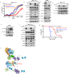
Fig. 6. Dependency on PHF6 is a specific consequence of SMARCB1 loss.
a Effects of shPHF6 vs shCTRL in SMARCB1-knockout 293 T cells treated with either GFP (+GFP) or SMARCB1 rescue (+SMARCB1) expression constructs. Data are represented from mean of n = 16 technical replicates per condition from one independent experiment. b Western blot analysis of histone modifications in 293TSMARCB1KO cells following knockdown of PHF6 and/or re-expression of SMARCB1 (48 h) or GFP (control) (n = 3 independent biological replicates). c Western blots of histone modifications in mouse embryonic fibroblasts derived from Phf6fl/y mice treated with either EGFP (control) or Cre to delete Phf6 (n = 2 independent biological replicates). Western blot analysis of the levels of Pol II Ser5P and Pol II Ser2P forms in (d) 293TSMARCB1KO following knockdown of PHF6 and/or re-expression of SMARCB1 or GFP (48 h) (n = 3 independent biological replicates) (e) in mouse embryonic fibroblasts derived from Phf6fl/fl mice treated with either EGFP (control) or Cre to delete Phf6 (n = 2 independent biological replicates). f Western blot analysis of SWI/SNF complex members in 293TSMARCB1KO cell line following knockdown of PHF6 and/or re-expression of SMARCB1 or GFP (48 h) (n = 3 independent biological replicates). g Kaplan–Meier survival curves demonstrating percent mice that are tumor free among cohorts of Phf6fl/y Smarcb1fl/fl Lck-Cre mice, Smarcb1fl/fl Lck-Cre mice, and Phf6fl/y Smarcb1fl/fl mice. n = 11 mice per group. Statistical analysis was performed using two-sided Mantel-Cox test; P = 0.0148. h Schematic illustration of PHF6 at the interface of chromatin (p300, H3K14ac modified nucleosome, ncBAF, and PBAF complexes) and transcriptional regulation (proximal promoter pause release; Pol II Ser5P). Figure 6h created with BioRender.com released under a Creative Commons Attribution-NonCommercial-NoDerivs 4.0 International license. Source data are provided as a Source Data file.

References

    1. Shain, A. H. & Pollack, J. R. The spectrum of SWI/SNF mutations, ubiquitous in human cancers. PLoS One8, e55119 (2013). 10.1371/journal.pone.0055119 - DOI - PMC - PubMed
    1. Kadoch, C. et al. Proteomic and bioinformatic analysis of mammalian SWI/SNF complexes identifies extensive roles in human malignancy. Nat. Genet45, 592–601 (2013). 10.1038/ng.2628 - DOI - PMC - PubMed
    1. Mittal, P. & Roberts, C. W. M. The SWI/SNF complex in cancer—biology, biomarkers and therapy. Nat. Rev. Clin. Oncol.17, 435–448 (2020). 10.1038/s41571-020-0357-3 - DOI - PMC - PubMed
    1. Versteege, I. et al. Truncating mutations of hSNF5/INI1 in aggressive paediatric cancer. Nature394, 203–206 (1998). 10.1038/28212 - DOI - PubMed
    1. Roberts, C. W., Galusha, S. A., McMenamin, M. E., Fletcher, C. D. & Orkin, S. H. Haploinsufficiency of Snf5 (integrase interactor 1) predisposes to malignant rhabdoid tumors in mice. Proc. Natl. Acad. Sci. USA97, 13796–13800 (2000). 10.1073/pnas.250492697 - DOI - PMC - PubMed

MeSH terms

Supplementary concepts