Review
. 2024 Aug 24;14(1):341.
doi: 10.1038/s41398-024-03047-y.

Applications of OPM-MEG for translational neuroscience: a perspective

Affiliations
Review

Applications of OPM-MEG for translational neuroscience: a perspective

Marion Brickwedde et al. Transl Psychiatry. .

Abstract

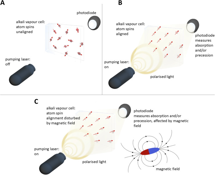
Magnetoencephalography (MEG) allows the non-invasive measurement of brain activity at millisecond precision combined with localization of the underlying generators. So far, MEG-systems consisted of superconducting quantum interference devices (SQUIDS), which suffer from several limitations. Recent technological advances, however, have enabled the development of novel MEG-systems based on optically pumped magnetometers (OPMs), offering several advantages over conventional SQUID-MEG systems. Considering potential improvements in the measurement of neuronal signals as well as reduced operating costs, the application of OPM-MEG systems for clinical neuroscience and diagnostic settings is highly promising. Here we provide an overview of the current state-of-the art of OPM-MEG and its unique potential for translational neuroscience. First, we discuss the technological features of OPMs and benchmark OPM-MEG against SQUID-MEG and electroencephalography (EEG), followed by a summary of pioneering studies of OPMs in healthy populations. Key applications of OPM-MEG for the investigation of psychiatric and neurological conditions are then reviewed. Specifically, we suggest novel applications of OPM-MEG for the identification of biomarkers and circuit deficits in schizophrenia, dementias, movement disorders, epilepsy, and neurodevelopmental syndromes (autism spectrum disorder and attention deficit hyperactivity disorder). Finally, we give an outlook of OPM-MEG for translational neuroscience with a focus on remaining methodological and technical challenges.


Conflict of interest statement

PJU reports having received research funding from Lilly UK and Lundbeck. RL reports personal fees from Medtronic. Prof. Kühn reports personal fees from Medtronic, and Boston Scientific. MH reports personal fees from Angelini, Bial, Desitin, Eisai, Jazz Pharma, UCB, and UNEEG within the last 3 years, outside the submitted work. All other authors report no biomedical financial interests or potential conflicts of interest.

Figures

Fig. 1
Fig. 1. OPM-Methodology.
A Alkali atoms are moving inside a vapor cell in a thermal random mixture of spin states. B ‘pumping’ the vapor cell with a laser producing polarized light induces transitions of most atoms into the same spin state. C The amount of light passing through the vapor cell becomes a function of the magnetic field, such as from brain activity, and can be measured with a photodiode.
Fig. 2
Fig. 2. OPM sensors and sensor arrays.
A Size of current generations of commercially available OPM sensors is as small as a USB stick (Quspin - https://quspin.com; Fieldline - https://fieldlineinc.com) (B) OPM sensors can be rigidly installed around a person’s head [146] (C), onto caps, similar to EEG electrodes [25] (Quspin - https://quspin.com) or (D) fitted into 3-D-printed helmets [147], (10.1111/nyas.14935). All arrangements can be adjusted for individual head sizes. No study participants are displayed in this figure and all material was edited with permission.
Fig. 3
Fig. 3. Comparison between OPM, EEG and SQUID-MEG.
A Comparison between SQUID-MEG and OPM-MEG for auditory evoked fields (n = 3 participants: S1, S2, S3) [30]. B Comparison between SQUID-MEG (blue line) and OPM-MEG (red line) for 80 trials of processing emotional (angry or happy) faces of source constructed M170 responses in 15 participants [148]. C Comparison of individual OPM-MEG and SQUID-MEG measurements during visual gamma responses for 6 comparable sensors (n = 1) [26]. D Comparison of auditory evoked fields (OPM-MEG) and potentials displaying typical differences between EEG and MEG-systems (n = 1) [24]. No changes were made to the original figure material, which was published under OPEN ACCESS license (CC BY 4.0; https://creativecommons.org/licenses/by/4.0/).
Fig. 4
Fig. 4. 40 Hz auditory steady-state responses during normal brain functioning and emerging psychosis.
A Average traces of six OPM sensors (top) and their time-frequency-analysis (bottom) over 250 trials [146]. Even without narrowband filtering, the 40 Hz response is clearly visible in each sensor (N = 22 healthy participants). B Inter-trial phase coherence, which illustrates the phase synchrony of the 40 Hz response over trials, and the difference of clinical groups from healthy controls (HC = 49 healthy controls; CHR-N = 38 participants with substance abuse and affective disorders; CHR-P = 116 participants with clinical high risk for psychosis; FEP = 33 participants with first episode psychosis). There is no difference between CHR-N and healthy controls, but the differences between CHR-P as well as FEP and healthy controls is apparent [62]. C The amplitude of the 40 Hz response in right Heschl’s Gyrus (auditory cortex) for the populations described in B. CHR-Ps as well as show a significantly reduced amplitude compared to healthy controls. No changes were made to the original figure material, which was published under OPEN ACCESS license (CC BY 4.0; https://creativecommons.org/licenses/by/4.0/).

References

    1. Kraguljac NV, McDonald WM, Widge AS, Rodriguez CI, Tohen M, Nemeroff CB. Neuroimaging biomarkers in Schizophrenia. Am J Psychiatry. 2021;178:509–21. 10.1176/appi.ajp.2020.20030340 - DOI - PMC - PubMed
    1. Uhlhaas PJ, Singer W. Neuronal dynamics and neuropsychiatric disorders: toward a translational paradigm for dysfunctional large-scale networks. Neuron. 2012;75:963–80. 10.1016/j.neuron.2012.09.004 - DOI - PubMed
    1. Fries P. Rhythms for cognition: communication through coherence. Neuron. 2015;88:220–35. 10.1016/j.neuron.2015.09.034 - DOI - PMC - PubMed
    1. Uhlhaas PJ, Singer W. Neural synchrony in brain disorders: relevance for cognitive dysfunctions and pathophysiology. Neuron. 2006;52:155–68. 10.1016/j.neuron.2006.09.020 - DOI - PubMed
    1. Uhlhaas PJ, Liddle P, Linden DEJ, Nobre AC, Singh KD, Gross J. Magnetoencephalography as a tool in psychiatric research: current status and perspective. Biol Psychiatry. 2017;2:235. - PMC - PubMed

LinkOut - more resources