. 2024 Aug 24;15(1):7294.
doi: 10.1038/s41467-024-51681-3.

The Chlamydia pneumoniae effector SemD exploits its host's endocytic machinery by structural and functional mimicry

Affiliations

The Chlamydia pneumoniae effector SemD exploits its host's endocytic machinery by structural and functional mimicry

Fabienne Kocher et al. Nat Commun. .

Abstract

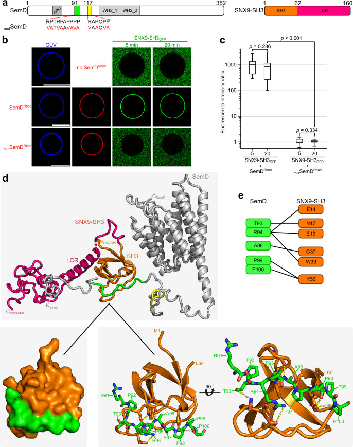
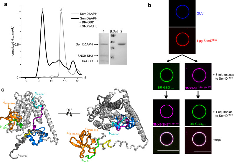
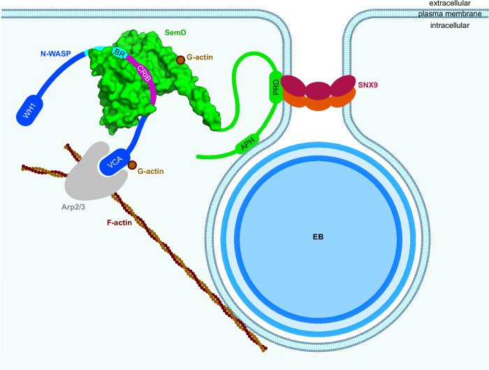
To enter epithelial cells, the obligate intracellular pathogen Chlamydia pneumoniae secretes early effector proteins, which bind to and modulate the host-cell's plasma membrane and recruit several pivotal endocytic host proteins. Here, we present the high-resolution structure of an entry-related chlamydial effector protein, SemD. Co-crystallisation of SemD with its host binding partners demonstrates that SemD co-opts the Cdc42 binding site to activate the actin cytoskeleton regulator N-WASP, making active, GTP-bound Cdc42 superfluous. While SemD binds N-WASP much more strongly than Cdc42 does, it does not bind the Cdc42 effector protein FMNL2, indicating effector protein specificity. Furthermore, by identifying flexible and structured domains, we show that SemD can simultaneously interact with the membrane, the endocytic protein SNX9, and N-WASP. Here, we show at the structural level how a single effector protein can hijack central components of the host's endocytic system for efficient internalization.


Conflict of interest statement

The authors declare no competing interests.

Figures

Fig. 1
Fig. 1. Crystal structure of SemDΔAPH.
a Schematic representation of the primary structure of SemD, containing an APH49-66, two proline-rich domains (PRD191-100, PRD2117-122) and two WH2 domains (WH2_1138-178, WH2_2179-216). SemDΔAPH67-382 is represented as a black bar. b The structure of SemDΔAPH as resolved by X-ray crystallography. The helices are depicted as cylinders and numbered from 1 to 9, starting at the N-terminus (α1- α9). In accordance with the colour code in a, the WH2_1 and WH2_2 are depicted in orange and red, respectively. The N-terminal E138 and the C-terminal E382 (both marked by black arrows) represent the first and last amino acids visible in the electron density. Right panel: 90° rotation. c SAXS best-fit CORAL model (χ2 value of 1.197), based on the SemDΔAPH crystal structure, and including the added flexible tails (further models are shown in Supplementary Fig. 1h). PRD1 and PRD2 are coloured in green and yellow, respectively. Right panel: 180° rotation. G67 is the N-terminal amino acid, while E382, the last C-terminal residue of SemD, is followed by the C-terminal 10x-His-Tag. d Electrostatic surface representation of SemDΔAPH highlighting the negatively (red) and positively (blue) charged patches. Right panel: 180° rotation.
Fig. 2
Fig. 2. SemD engages with BR-GBD in a Cdc42GTP-mimicking manner.
a Schematic representation of the N-WASP primary sequence. BR-GBD142-273 was used for complex formation with SemDΔAPH. It contains the BR181-197 domain (basic region, cyan), the CRIB198-213 domain (Cdc42/Rac interactive binding motif, magenta) and the C-sub214-250 domain (blue). b The structure of SemDΔAPH in complex with BR-GBD as resolved by X-ray crystallography, shown in cartoon representation. SemDΔAPH is shown in light grey, BR-GBD is coloured in dark grey with the BR domain in cyan, the CRIB domain in magenta and C-sub domain in blue. The zoom in shows details of the binding of SemDΔAPH to BR-GBD. Important residues of SemDΔAPH and BR-GBD are shown in stick representation, while the rest of SemDΔAPH is shown as cartoon. Interactions (<3.5 Å) are shown by the yellow dashes. c Schematic representation of the detailed interactions between SemDΔAPH and BR-GBD. d Electrostatic representation of SemDΔAPH, highlighting the negatively charged patch in red and positively charged surface areas in blue. BR-GBD is coloured in dark grey (cartoon) with the BR domain in cyan and the CRIB domain in magenta, both depicted with stick residues.
Fig. 3
Fig. 3. SemD recruits the BR-GBD region of N-WASP to membrane vesicles.
a Amino acid sequence of WASP219-259 (human) and N-WASP181-221 (Rattus norvegicus) (of which the latter is identical to N-WASP184-224 from human) (top). The orange box shows the sequence involved in binding to Cdc42GTP and SemD, respectively. The lower panel shows the structures of Cdc42GTP in complex with BR-GBDWASP (PDB: 1CEE) and SemDΔAPH bound to BR-GBDN-WASP. Cdc42GTP binds to the positively charged C-terminal KKK230-232 motif of the BR from WASP and embeds the CRIB domain in the less charged binding groove. The same binding mechanism is observed for SemD, in which a negatively charged patch engages with the positively charged KKR192-194 motif within the N-WASP BR domain and inserts the subsequent N-WASP CRIB domain into the SemD binding groove. b Schematic representation of BR-GBD and the deletion variants BR-GBDΔ (lacking the BR175-197 domain) and BR-GBDΔΔ (lacking the BR175-197 and the CRIB198-213 domains). Dashed lines in square brackets mark the deleted protein regions. The first and last deleted amino acids are indicated. c Confocal images of PS-containing GUVs with rhodamine-labelled SemD (SemDRhod) and BR-GBD variants fused to GFP (BR-GBDGFP). (scale bars 10 µm). d Quantification of bound protein to the GUV membrane based on the ratio of the maximal fluorescence at the perimeter of the GUV to the average background fluorescence outside the GUV. For each variant and time point, the fluorescence intensity ratio was calculated for up to 22 independent GUVs. The data are represented as boxplots with whiskers. The boxes are limited by the 25th and 75th percentile, including 50% of the data. The centre line shows the mean score. Whiskers denote 5–95% of all data and outliers are shown as grey dots. For comparing two groups, an unpaired, two-sided Student’s t test was used. The data are representative for two independent data sets, both yielding similar results. Source data of both data sets are provided as Source Data file.
Fig. 4
Fig. 4. Relative to Cdc42GppNHp, SemDΔAPH displays enhanced and specific binding to N-WASP.
a GFP Trap® Pulldown experiments using equimolar ratios of purified recombinant SemDΔAPHHis, and active Cdc42 bound to the non-hydrolysable GTP analogue (Cdc42GppNHp), were used to test their respective binding to BR-GBDGFP-His. Complex formation between BR-GBDGFP-His and SemDΔAPHHis or Cdc42GppNHp served as positive controls. Flow through (FT), wash 6 (W6) and elution (El.) fractions were analysed by SDS/PAGE and probed with anti-His (SemDΔAPHHis and BR-GBDGFP-His) and anti-Cdc42 (Cdc42) antibodies. Pulldown experiments were repeated three times with similar results. Replicates and negative controls are provided in Supplementary Fig. 4a and as Source Data to Fig. 4. b Quantification of western blotting in a is described in methods. Normalised (norm.) data are displayed as mean ± s.d. (n = 3 biologically independent experiments). Unpaired, two-sided Student’s t-test was used to compare two groups. c, d Stopped-Flow experiments used to test the binding of equimolar ratios of BR-GBDHis and Cdc42mGppNHp (c), as well as the displacement of Cdc42mGppNHp from BR-GBDHis by the addition of an equimolar amount of SemDΔAPHHis (d). The lower panels schematically indicate the relevant interactions. e GST-Agarose pulldown experiments used to probe the interactions of Formin L2GST (FMNL2GST) with Cdc42GppNHp and SemDΔAPHHis with Cdc42GppNHp. Complex formation between Cdc42GppNHp and FMNL2GST served as a positive control. Flow through (FT), wash 6 (W6) and elution (El.) fractions were analysed by SDS/PAGE and probed with anti-His (SemDΔAPHHis), anti-GST (FMNL2GST) and anti-Cdc42 (Cdc42) antibodies. Pulldown experiments were repeated three times with similar results. Replicates and negative controls are provided in Supplementary Fig. 4b, c and as Source Data to Fig. 4. f Quantification of western blotting in e is described in methods. Normalised (norm.) data are displayed as mean ± s.d. (n = 3 biologically independent experiments). Unpaired, two-sided Student’s t test was used to compare two groups.
Fig. 5
Fig. 5. SemD binds to SNX9-SH3 PRD1.
a (left) Schematic representation of SemD with PRD1 and PRD2 in green and yellow, respectively. Mutations for SemDmut are indicated by the amino acids in red. (right) Schematic representation of SNX9-SH3 with the SH3 in orange and the low complexity region (LCR) in pink. b Confocal images of PS-containing GUVs with labelled SemD or SemDmut and SNX9-SH3GFP. (scale bars 10 µm). c Quantification of bound protein to the GUV membrane based on the ratio of the maximal fluorescence at the perimeter of the GUV to the average background fluorescence outside the GUV. For each variant and time point, the fluorescence intensity ratio was calculated for up to four independent GUVs. The data are represented as boxplots with whiskers. The boxes are limited by the 25th and 75th percentile, including 50 % of the data. The centre line shows the mean score. Whiskers denote 5–95% of all data and outliers are shown as grey dots. For comparing two groups, an unpaired, two-sided Student’s t test was used. The data are representative for three independent data sets, all yielding similar results. Source data of all data sets are provided as Source Data file. d Cartoon model of SemDΔAPH (grey) with SNX9-SH3 (SH3 in orange) as determined by SAXS using the programme CORAL. SNX9-SH3 (orange) binds to PRD1 (green) of SemDΔAPH. (zoom in) The interactions between the two domains are displayed. e Proposed details of the interactions between the two domains, based on the model shown in d.
Fig. 6
Fig. 6. SemD simultaneously interacts with various binding partners.
a SEC chromatograms of the complex composed of SemDΔAPH, BR-GBD and SNX9-SH3 (black), or SemDΔAPH alone (grey). The absorbance at 280 nm was normalised for the maximal absolute absorbance of the individual sample. The chromatogram of the complex revealed a major peak eluting at 9.9 ml (peak 1), while SemDΔAPH alone elutes at 14.2 ml (peak 2). The protein compositions of peaks 1 and 2 were analysed on an SDS gel (right) after staining with Coomassie brilliant blue. Lanes 1 and 2 were loaded with samples of the indicated peaks (n = 1). b Confocal images of PS-containing GUVs incubated with SemDRhod. A three-fold excess of either BR-GBDGFP (n = 4) or labelled SNX9-SH3 (SNX9-SH3DyLight 650) (n = 6) was added, before the third binding partner was added in an equimolar ratio to SemD (scale bars 10 µm). c The structures of SNX9-SH3 and BR-GBD obtained by SAXS overlaid on SemDΔAPH. The nine core helices of SemDΔAPH are depicted in grey and the PRD1 and PRD2 in green and yellow, respectively. BR-GBD is depicted in cyan, magenta and blue, in accordance with the colour scheme in Fig. 2, and the depiction of the SNX9-SH3 domain in orange follows the colour scheme used in Fig. 5. Note that the three-dimensional orientation of the bound SH3 domain towards the nine-helix core might be different, owing to the presence of the flexible linker in between the two. Right panel: 90° rotation.
Fig. 7
Fig. 7. A chlamydial effector exploits structural and functional mimicry to manipulate the host endocytic machinery.
The Cpn elementary body (EB) secrets SemD into the host cell, which binds to the inner leaflet of the plasma membrane. There, SemD recruits, binds and activates N-WASP by structurally and functionally mimicking the Cdc42GTP activation mechanism. SemD interacts with the C-terminal, positively charged amino acids of the N-WASP BR domain and further, the CRIB domain binds into the SemD binding groove. This then leads to the release of N-WASP from its auto-inhibited state. SemD also binds to the SNX9-SH3 domain, which brings the SNX9-BAR domain closer to the membrane. This in turn induces membrane deformation and eventually leads to closure of the matured endocytic vesicle. Due to the arrangement of the individual binding domains, which are connected by flexible linker regions, the binding sites can be freely oriented in 3D space, thus minimising steric hindrance. This can explain why SemD is postulated to be capable of binding simultaneously to the PM, SNX9 and N-WASP in vivo and hijacking their functions to promote the growth and maturation of the endocytic vesicle.

References

    1. Kuo, C. C. et al. Chlamydia pneumoniae (TWAR) in coronary arteries of young adults (15-34 years old). Proc. Natl Acad. Sci. USA92, 6911–6914 (1995). 10.1073/pnas.92.15.6911 - DOI - PMC - PubMed
    1. Hahn, D. L. Chlamydia pneumoniae and chronic asthma: updated systematic review and meta-analysis of population attributable risk. PLoS ONE16, e0250034 (2021). 10.1371/journal.pone.0250034 - DOI - PMC - PubMed
    1. Balin, B. J. et al. Chlamydia pneumoniae: an etiologic agent for late-onset dementia. Front. Aging Neurosci.10, 302 (2018). 10.3389/fnagi.2018.00302 - DOI - PMC - PubMed
    1. Beagley, K. W., Huston, W. M., Hansbro, P. M. & Timms, P. Chlamydial infection of immune cells: altered function and implications for disease. Crit. Rev. Immunol.29, 275–305 (2009). 10.1615/CritRevImmunol.v29.i4.10 - DOI - PubMed
    1. Gerard, H. C. et al. Chlamydophila (Chlamydia) pneumoniae in the Alzheimer’s brain. FEMS Immunol. Med. Microbiol.48, 355–366 (2006). 10.1111/j.1574-695X.2006.00154.x - DOI - PubMed

MeSH terms

LinkOut - more resources