

Imaging in pelvic exenteration-a multidisciplinary practice guide from the ESGAR-SAR-ESUR-PelvEx collaborative group
- PMID: 39181949
- PMCID: PMC12021987
- DOI: 10.1007/s00330-024-10940-z
Imaging in pelvic exenteration-a multidisciplinary practice guide from the ESGAR-SAR-ESUR-PelvEx collaborative group
Erratum in
-
Correction: Imaging in pelvic exenteration-a multidisciplinary practice guide from the ESGAR-SAR-ESUR-PelvEx collaborative group.Eur Radiol. 2025 Sep;35(9):5870-5871. doi: 10.1007/s00330-025-11437-z. Eur Radiol. 2025. PMID: 40063109 Free PMC article. No abstract available.
Abstract
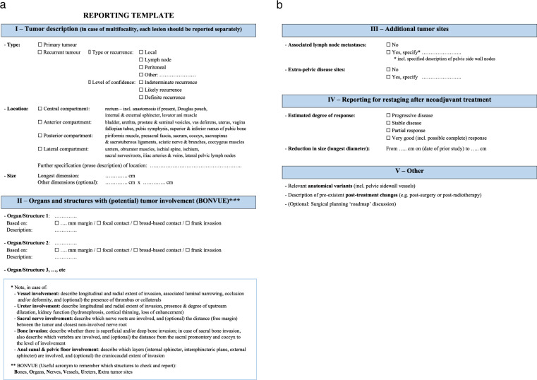
Pelvic exenteration (PE) is a radical surgical approach designed for the curative treatment of advanced pelvic malignancies, requiring en-bloc resection of multiple pelvic organs. While the procedure is radical, it has shown promise in enhancing long-term survival and is now comparable in surgical mortality to elective resections for primary pelvic cancers. Imaging plays a crucial role in preoperative planning, with MRI, CT, and PET/CT being pivotal in assessing the extent of cancer and formulating a surgical roadmap. This paper presents clinical practice guidelines for imaging in the context of PE, developed jointly by ESGAR, SAR, ESUR, and the PelvEx Collaborative. These guidelines aim to standardize imaging protocols and reporting to improve the preoperative assessment and facilitate decision-making in the multidisciplinary treatment of pelvic cancers. Our recommendations underscore the importance of a multidisciplinary approach and the need for clear and precise imaging reports to optimize patient care. CLINICAL RELEVANCE STATEMENT: Our recommendations underscore the importance of a multidisciplinary approach and the need for clear and precise imaging reports to optimize patient care. KEY POINTS: MRI is mandatory for local staging in pelvic exenteration. Structured reporting (using the template provided in this guide) is recommended. Multidisciplinary review of imaging is critical for surgical planning.
Keywords: CT; Cancer; MRI; PET/CT; Pelvic exenteration.
© 2024. The Author(s).
Conflict of interest statement
Compliance with ethical standards. Guarantor: The scientific guarantor of this publication is S.N. Conflict of interest: The authors of this manuscript declare no relationships with any companies whose products or services may be related to the subject matter of the article. Statistics and biometry: No complex statistical methods were necessary for this paper. Informed consent: Written informed consent was waived by the Institutional Review Board. Ethical approval: Institutional Review Board approval was not required because of the nature of the article. Study subjects or cohorts overlap: None. Methodology: Recommendations
Figures

References
-
- Egger EK, Liesenfeld H, Stope MB et al (2021) Pelvic exenteration in advanced gynecologic malignancies—who will benefit? Anticancer Res 41:3037–3043 - PubMed
-
- Gould LE, Pring ET, Drami I et al (2022) A systematic review of the pathological determinants of outcome following resection by pelvic exenteration of locally advanced and locally recurrent rectal cancer. Int J Surg 104:106738 - PubMed
-
- PelvEx C (2023) A review of functional and surgical outcomes of gynaecological reconstruction in the context of pelvic exenteration. Surg Oncol 52:101996 - PubMed
-
- PelvExCollaborative (2019) Pelvic exenteration for advanced nonrectal pelvic malignancy. Ann Surg 270:899–905 - PubMed
Publication types
MeSH terms
Grants and funding
LinkOut - more resources
Full Text Sources
Research Materials
Miscellaneous
