. 2024 Aug 24;30(1):130.
doi: 10.1186/s10020-024-00900-0.

Antibiotic-induced gut microbiota disruption promotes vascular calcification by reducing short-chain fatty acid acetate

Affiliations

Antibiotic-induced gut microbiota disruption promotes vascular calcification by reducing short-chain fatty acid acetate

Shi-Yu Zeng et al. Mol Med. .

Abstract

Background: Vascular calcification is a common vascular lesion associated with high morbidity and mortality from cardiovascular events. Antibiotics can disrupt the gut microbiota (GM) and have been shown to exacerbate or attenuate several human diseases. However, whether antibiotic-induced GM disruption affects vascular calcification remains unclear.

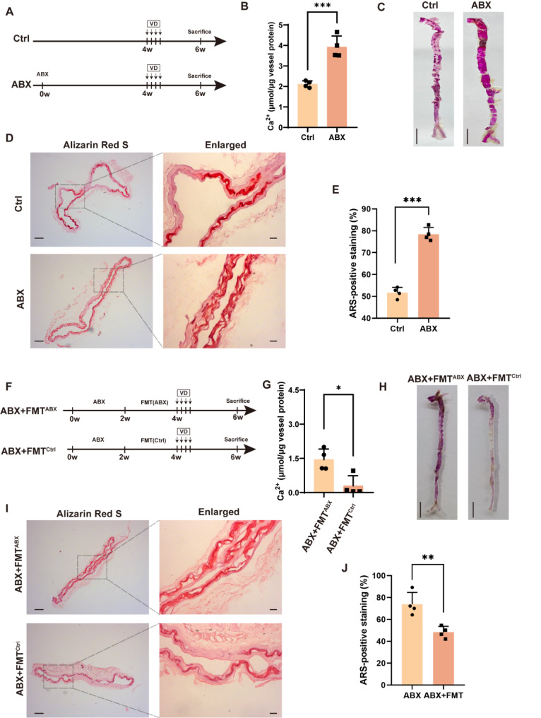
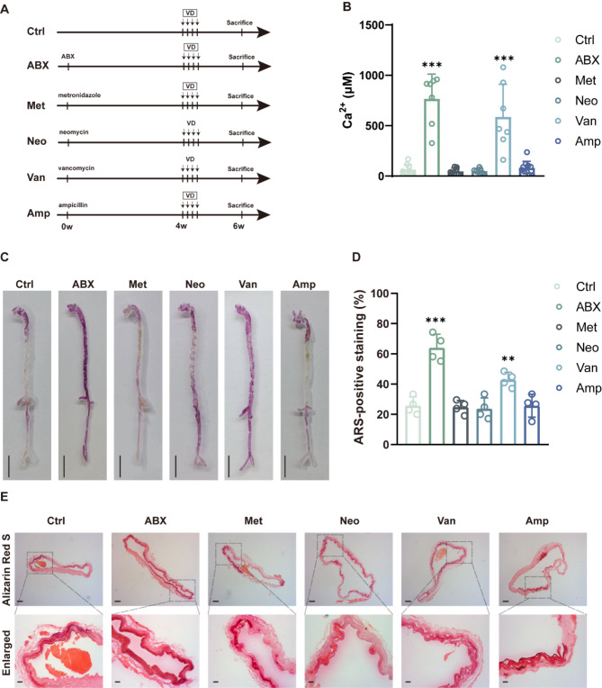
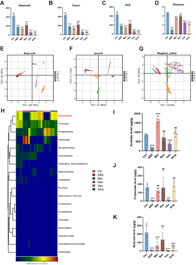
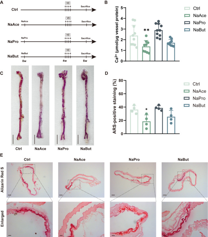
Methods: Antibiotic cocktail (ABX) treatment was utilized to test the potential effects of antibiotics on vascular calcification. The effects of antibiotics on GM and serum short-chain fatty acids (SCFAs) in vascular calcification mice were analyzed using 16 S rRNA gene sequencing and targeted metabolomics, respectively. Further, the effects of acetate, propionate and butyrate on vascular calcification were evaluated. Finally, the potential mechanism by which acetate inhibits osteogenic transformation of VSMCs was explored by proteomics.

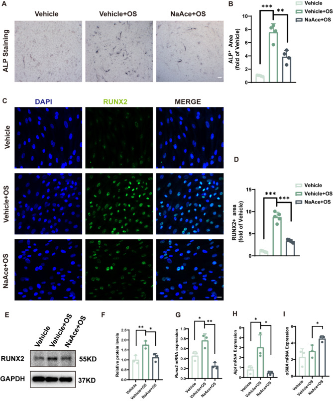
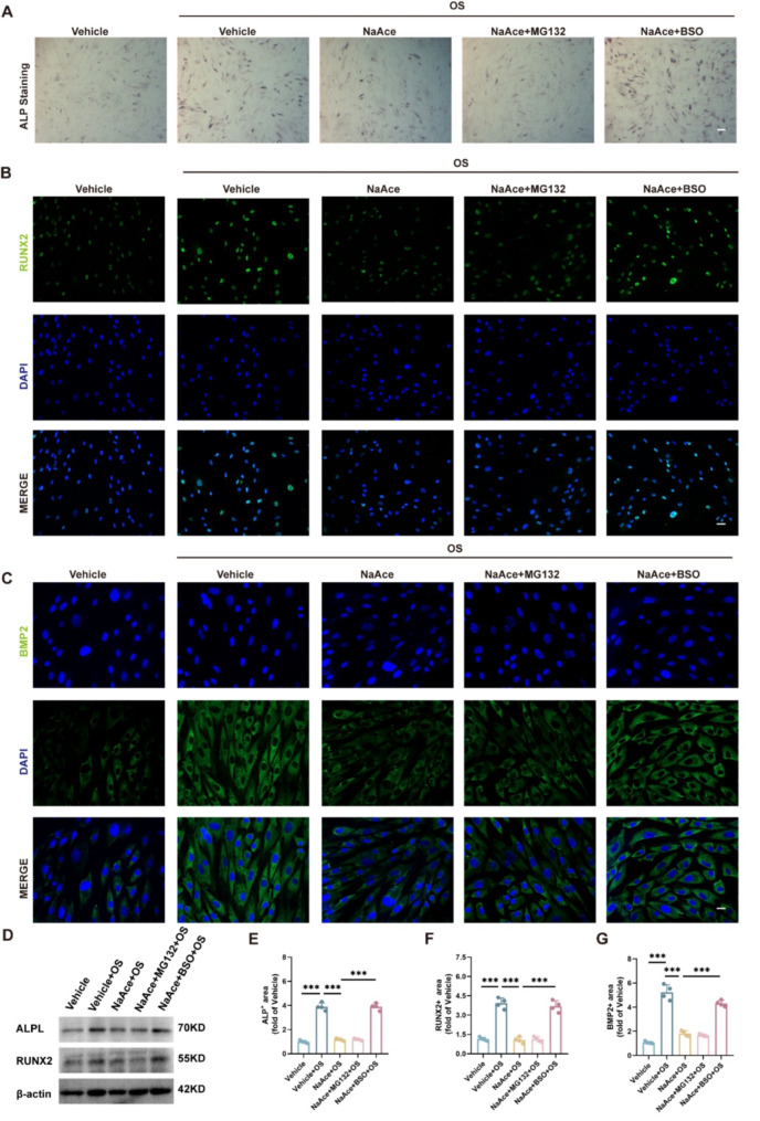
Results: ABX and vancomycin exacerbated vascular calcification. 16 S rRNA gene sequencing and targeted metabolomics analyses showed that ABX and vancomycin treatments resulted in decreased abundance of Bacteroidetes in the fecal microbiota of the mice and decreased serum levels of SCFAs. In addition, supplementation with acetate was found to reduce calcium salt deposition in the aorta of mice and inhibit osteogenic transformation in VSMCs. Finally, using proteomics, we found that the inhibition of osteogenic transformation of VSMCs by acetate may be related to glutathione metabolism and ubiquitin-mediated proteolysis. After adding the glutathione inhibitor Buthionine sulfoximine (BSO) and the ubiquitination inhibitor MG132, we found that the inhibitory effect of acetate on VSMC osteogenic differentiation was weakened by the intervention of BSO, but MG132 had no effect.

Conclusion: ABX exacerbates vascular calcification, possibly by depleting the abundance of Bacteroidetes and SCFAs in the intestine. Supplementation with acetate has the potential to alleviate vascular calcification, which may be an important target for future treatment of vascular calcification.

Keywords: Acetate; Antibiotic; Gut microbiota; Short-chain fatty acid; Vancomycin; Vascular calcification.


Conflict of interest statement

The authors declare no competing interests.

Figures

Fig. 1
Fig. 1
Antibiotic cocktail treatment exacerbates VD-induced vascular calcification in mice by modulation of GM. (A) Flow diagram of the antibiotic cocktail (ABX) intervention experiment. (B) Quantitative evaluation of aortic calcium content in mice. n = 4 per group. (C) Macroscopic observation of arterial vascular calcification. Scale bar = 0.5 cm. (D) Calcification of ascending aortic vessels based on alizarin red-stained tissue sections. Scale bar = 100 μm (right) and 20 μm (left). (E) Relative quantification of positive ARS area in ascending aortic vascular sections. n = 4 per group. (F) A flow diagram of the fecal microbiota transplantation (FMT) experiment. (G) Quantitative evaluation of aortic calcium content in mice. n = 4 per group. (H) Macroscopic observation of arterial vascular calcification. Scale bar = 0.5 cm. (I) Calcification of ascending aortic vessels based on alizarin red-stained tissue sections. Scale bar = 100 μm (right) and 20 μm (left). (J) Relative quantification of positive ARS area in ascending aortic vascular sections. n = 4 per group. Data are presented as mean ± SD. *P < 0.05, **P < 0.01, and ***P < 0.001
Fig. 2
Fig. 2
Vancomycin treatment exacerbates VD-induced vascular calcification in mice. (A) Flow diagram of the single antibiotic intervention experiment. (B) Quantitative evaluation of aortic calcium content in mice. n = 7–10 per group. (C) Macroscopic observation of arterial vascular calcification. Scale bar = 0.5 cm. (D) Relative quantification of positive ARS area in ascending aortic vascular sections. n = 4 per group. (E) Calcification of ascending aortic vessels based on alizarin red-stained tissue sections. Scale bar = 100 μm (top) and 20 μm (bottom). Data are presented as mean ± SD. *P < 0.05, **P < 0.01, and ***P < 0.001
Fig. 3
Fig. 3
Antibiotic treatment alters the structure and composition of GM and reduces the production of GM metabolite SCFAs. (A to D) Observed number of OTUs and estimated OTU richness (Chao1, ACE, and Shannon) in fecal microbiota of mice. n = 5 per group. (E to G) Principal coordinate analysis (PCoA) plot based on Braycurtis distances, Jaccard distances and Weighted_unifrac distances. n = 5 per group. (H) Relative abundance of the identified fecal microbiota at the phylum level as detected by 16 S rRNA gene sequencing. n = 5 per group. (I to J) short-chain fatty acids in mouse serum. n = 5 per group. Data are presented as mean ± SD. *P < 0.05, **P < 0.01, and ***P < 0.001
Fig. 4
Fig. 4
NaAce supplementation alleviates VD-induced vascular calcification in mice. (A) Flow diagram of the short-chain fatty acids intervention experiment. (B) Quantitative evaluation of aortic calcium content in mice. n = 9–10 per group. (C) Macroscopic observation of arterial vascular calcification. Scale bar = 0.5 cm. (D) Relative quantification of positive ARS area in ascending aortic vascular sections. n = 4 per group. (E) Calcification of ascending aortic vessels based on alizarin red-stained tissue sections. Scale bar = 100 μm (top) and 20 μm (bottom). Data are presented as mean ± SD. *P < 0.05, **P < 0.01, and ***P < 0.001
Fig. 5
Fig. 5
NaAce inhibits osteoblast differentiation of VSMCs in vitro. (A) Alkaline phosphatase (ALP) staining images of VSMCs. Scale bar = 200 μm. (B) Relative quantification of ALP activity. n = 4 per group. (C) Immunofluorescence images of Runx2 protein in VSMCs. Scale bar = 20 μm. (D) Quantification of relative expression of Runx2 protein in VSMCs. n = 5 per group. (E and F) Protein expression of Runx2 determined using western blotting. n = 3 per group. (G to I) Runx2, Alpl, and αSMA mRNA expression in VSMCs. n = 3 per group. Data are presented as mean ± SD. *P < 0.05, **P < 0.01, and ***P < 0.001
Fig. 6
Fig. 6
Proteomic analysis of NaAce-treated and non-treated VSMCs. (A) Cluster tree for detecting reproducibility of samples in three groups. (B) Volcano map showing proteins undergoing up- or down-regulation in OS-induced VSMCs by NaAce intervention. (C) Up- or down-regulated proteins resulting from NaAce treatment in terms of subcellular localization GO annotations. (D) GO enrichment analysis of differential proteins in the OS + NaAce group relative to the OS group. (E) KEGG enrichment analysis of differential proteins in the OS + NaAce group relative to the OS group. n = 3 per group. Data are presented as mean ± SD
Fig. 7
Fig. 7
Glutathione metabolism may be involved in the inhibitory effect of NaAce on osteogenesis. (A) Alkaline phosphatase (ALP) staining images of VSMCs. Scale bar = 200 μm. (B) Immunofluorescence images of Runx2 protein in VSMCs. Scale bar = 20 μm. (C) Immunofluorescence images of BMP2 protein in VSMCs. Scale bar = 20 μm. (D) Protein expression of Runx2 and ALPL determined using western blotting. n = 3 per group. (E) Relative quantification of ALP activity. n = 4 per group. (F) Quantification of relative expression of Runx2 positive area. n = 4 per group. (G) Quantification of relative expression of BMP2 positive area. n = 4 per group. Data are presented as mean ± SD. *P < 0.05, **P < 0.01, and ***P < 0.001
Fig. 8
Fig. 8
Schematic diagram. Short-chain fatty acid reduction in antibiotic-induced intestinal dysbiosis aggravates vascular calcification

References

    1. Akram F, Imtiaz M, Haq IU. Emergent crisis of antibiotic resistance: a silent pandemic threat to 21(St) century. Microb Pathog. 2023;174:105923. 10.1016/j.micpath.2022.105923 - DOI - PubMed
    1. Aoki R, Onuki M, Hattori K, Ito M, Yamada T, Kamikado K, Kim YG, Nakamoto N, Kimura I, Clarke JM, Kanai T, Hase K. Commensal microbe-derived acetate suppresses NAFLD/NASH development via hepatic FFAR2 signalling in mice. Microbiome. 2021;9:188. 10.1186/s40168-021-01125-7 - DOI - PMC - PubMed
    1. Armet AM, Deehan EC, O’Sullivan AF, Mota JF, Field CJ, Prado CM, Lucey AJ, Walter J. Rethinking healthy eating in light of the gut microbiome. Cell Host Microbe. 2022;30:764–85. 10.1016/j.chom.2022.04.016 - DOI - PubMed
    1. Browne AJ, Chipeta MG, Haines-Woodhouse G, Kumaran EPA, Hamadani BHK, Zaraa S, Henry NJ, Deshpande A, Reiner RC Jr., Day NPJ, Lopez AD, Dunachie S, Moore CE, Stergachis A, Hay SI, Dolecek C. Global antibiotic consumption and usage in humans, 2000-18: a spatial modelling study. Lancet Planet Health. 2021;5:e893–904. 10.1016/S2542-5196(21)00280-1 - DOI - PMC - PubMed
    1. Chambers LM, Esakov Rhoades EL, Bharti R, Braley C, Tewari S, Trestan L, Alali Z, Bayik D, Lathia JD, Sangwan N, Bazeley P, Joehlin-Price AS, Wang Z, Dutta S, Dwidar M, Hajjar A, Ahern PP, Claesen J, Rose P, Vargas R, Brown JM, Michener CM, Reizes O. Disruption of the gut microbiota confers Cisplatin Resistance in Epithelial Ovarian Cancer. Cancer Res. 2022;82:4654–69. 10.1158/0008-5472.CAN-22-0455 - DOI - PMC - PubMed

MeSH terms

LinkOut - more resources