. 2024 Sep-Oct:105:107385.
doi: 10.1016/j.ntt.2024.107385. Epub 2024 Aug 23.

Differences in withdrawal symptoms, microglia activity, and cognitive functioning in rats exposed to continuous low-dose heroin in-utero

Affiliations

Differences in withdrawal symptoms, microglia activity, and cognitive functioning in rats exposed to continuous low-dose heroin in-utero

Sara L Mills-Huffnagle et al. Neurotoxicol Teratol. 2024 Sep-Oct.

Abstract

Introduction: Opioid use during pregnancy and subsequent neonatal opioid withdrawal syndrome (NOWS) have been associated with poor developmental outcomes including cognitive functioning. Less is known about the underlying molecular effects of prenatal opioid exposure and subsequent withdrawal; however, given the recent increase in NOWS cases, there is a pressing need to better understand these effects, which may partially explain cognitive deficits that have been observed in both preclinical NOWS models and patients with NOWS. This study evaluated the effects of prenatal heroin exposure and subsequent precipitated withdrawal symptoms on microglial reactivity in the nucleus accumbens (NAc), dorsal hippocampus (HC), and ventral tegmental area (VTA) in rat neonates, as well as cognitive functioning at three developmental time points using the Morris Water Maze (MWM) task.

Methods: Heroin or saline (2 mg/kg) was randomly assigned and administered to six pregnant Sprague Dawley rat dams via osmotic minipump. A total of 63 rat neonates underwent naloxone-precipitated (5 mg/kg, subcutaneous injection) withdrawal testing at postnatal day 10 (PN10). Following withdrawal testing, neonates were randomly assigned to undergo perfusion and subsequent immunohistochemistry experiments to fluoresce Iba-1 for microglia detection, or to undergo the MWM task at three separate developmental time points (PN21-23; PN37; PN60) for cognitive testing.

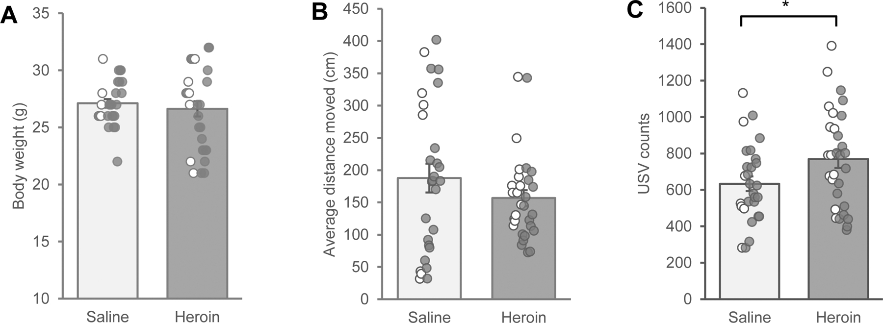
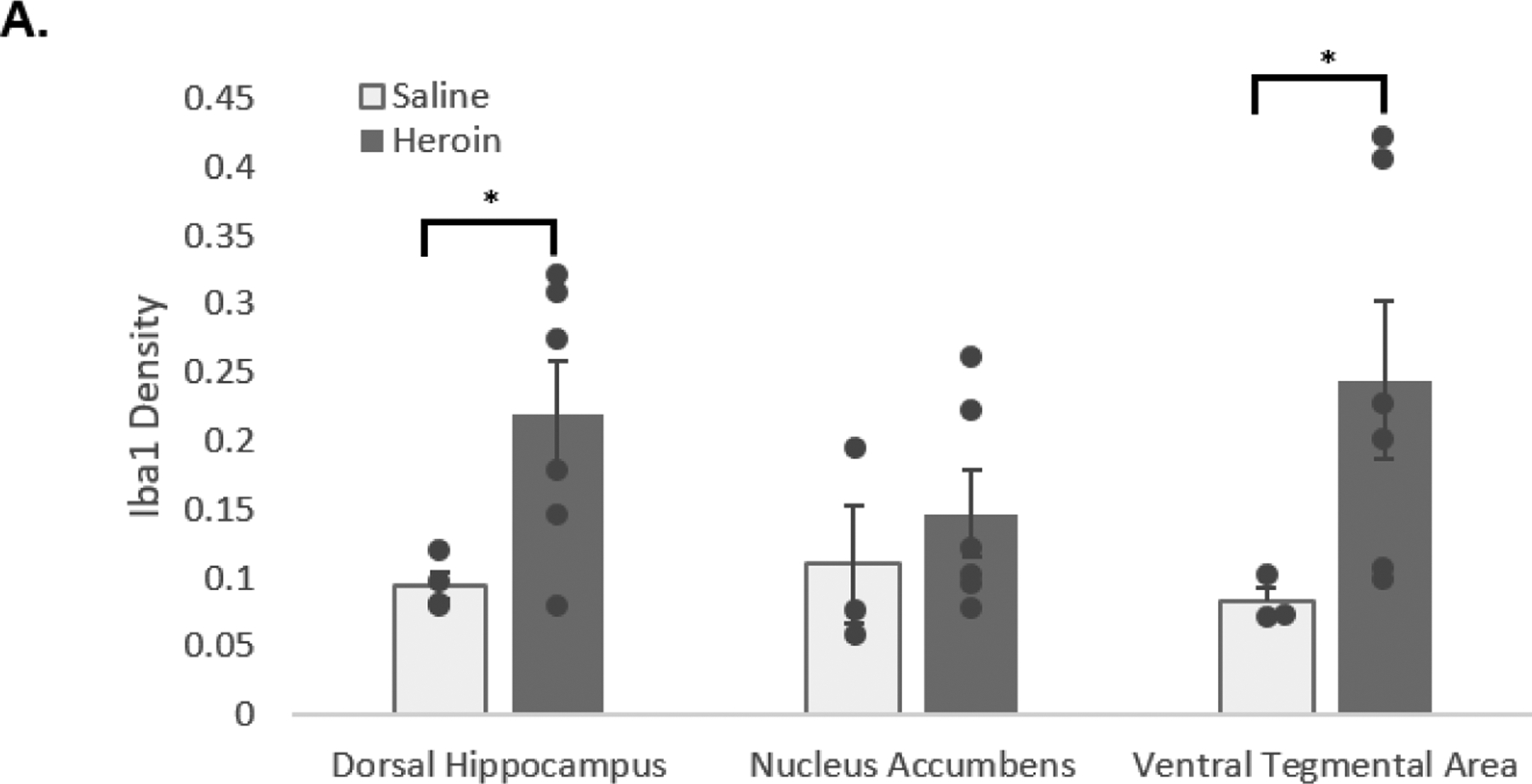
Results: Results suggest that in-utero heroin exposure led to an increase in ultrasonic vocalizations during naloxone-precipitated withdrawal; a sensitive index of withdrawal in rat neonates. Additional results suggest increased microglial reactivity in the HC and VTA, but not the NAc, as well as reduced performance during the MWM in the group exposed to heroin in-utero.

Discussion: Together, these data suggest that in-utero opioid exposure is associated with microglial reactivity in brain regions associated with learning and memory, and may be associated with later cognitive deficits. Further research is needed to characterize these findings, which may inform future therapeutic strategies for this vulnerable population.

Keywords: Cognitive functioning; Microglia reactivity; Neonatal opioid withdrawal syndrome; Prenatal heroin exposure.


Conflict of interest statement

Declaration of competing interest The authors declare that they have no known competing financial interests or personal relationships that could have appeared to influence the work reported in this paper. Sara L. Mills-Huffnagle, MS reports financial support was provided by National Institutes of Health. Jennifer E. Nyland, PhD reports financial support was provided by National Institute of Health. If there are other authors, they declare that they have no known competing financial interests or personal relationships that could have appeared to influence the work reported in this paper.

Figures

Figure 1.
Figure 1.. Timeline of experimental procedures.
At E14, a saline- or heroin-primed osmotic minipump was placed in the pregnant rat dams. At PN10, saline- or heroin-exposed rat pups underwent naloxone-precipitated withdrawal, and locomotor activity and ultrasonic vocalizations were recorded. Afterward, 10 rat pups underwent transcardiac perfusion for collection of brain tissue and subsequent immunohistochemistry (IHC) experiments, and 14 rat pups completed the Morris Water Maze test at PN23, PN37, and PN60.
Figure 2:
Figure 2:. Holding apparatus for withdrawal testing and recording.
These images display the holding apparatus for the rat neonates during withdrawal testing, as well as EthovisionXT®’s tracking procedures over time. The apparatus is approximately 7 ¾” L x 11 ⅜” W x 5” H. Recording occurred through a Basler Ace monochrome camera with Gigabit Ethernet interface and tracking activity through the associated EthovisionXT® software (Noldus). Tracking activity is indicated by the red line, which in turn allows for the exact measurement of the total distance moved in centimeters (cm).
Figure 3:
Figure 3:. Assessment of neonatal opioid withdrawal at PN10.
A) Mean body weight (±SEM): There was no difference in weight between the saline-exposed and heroin-exposed rats at PN10. There was an emerging trend in weight differences at PN10 between heroin-exposed male (open circles) and female (closed circles) rats. B) Average distance moved (±SEM): Saline-exposed rats appeared to move more compared with heroin-exposed rats; however, this did not reach significance. C) Mean USVs (±SEM): Heroin-exposed rats had more USV calls compared with saline-exposed rats (p < 0.05). USV = ultrasonic vocalizations. Saline n = 28 (7 male, 21 female); Heroin n = 30 (13 male, 17 female). * p < 0.05
Figure 4:
Figure 4:. Iba1 densitometry.
A) Mean density of Iba1 labeled cells (±SEM): Heroin-exposed rats exhibited higher levels of Iba1 density in the dorsal hippocampus (HC) at PN10 when compared with the saline-exposed rats (saline n = 4; heroin n = 6). The heroin-exposed rats also exhibited slightly higher levels of Iba1 density in the nucleus accumbens (NAc) at PN10 when compared with the saline-exposed rats, but this was not significant (saline n = 3; heroin n = 6). The heroin-exposed rats did have significantly higher levels of Iba1 density in the ventral tegmental area (VTA) at PN10 when compared with saline-exposed rats (saline n = 3; heroin n = 6). B) DAPI-stained sample images to show regions of interest including the HC, NAc, and VTA, as well as immunofluorescent image samples of each area with Iba1 microglia labeling in a saline-exposed (left) and heroin-exposed rat (right). Iba1 = Ionized calcium-binding adapter molecule 1. *p < 0.05; LV = lateral ventricle; PAG = periaqueductal gray; IP = interpenduncular nucleus; SNr = substania nigra
Figure 5:
Figure 5:. Cognitive assessments using the Morris Water Maze (MWM).
A) Mean time to complete the MWM task (±SEM): There was no difference in the amount of time (in seconds) to complete the MWM task at PN21–23, PN37, or PN60 between the saline-exposed and heroin-exposed rats (F (1,12) = 0.223, p = 0.646). B) Mean number of crossovers during the MWM testing sessions (±SEM): Heroin-exposed rats had fewer crossovers during the MWM test compared with the saline-exposed rats at timepoints PN23 and PN37, but not at PN60. C) Mean time spent in the original location of the platform during the MWM testing sessions (±SEM): Heroin-exposed rats spent less time in the general area of the platform after its removal during the MWM testing sessions compared with the saline-exposed rats. This trend reached significance at PN37 and PN60, but not at PN23. Saline n = 6; Heroin n=8 * p < 0.05, **p < 0.001

References

    1. Amalric M, Koob GF. Low doses of methylnaloxonium in the nucleus accumbens antagonize hyperactivity induced by heroin in the rat. Pharmacol Biochem Behav. 1985. Sep;23(3):411–5. - PubMed
    1. Andersen JM, Høiseth G, Nygaard E. (2020). Prenatal exposure to methadone or buprenorphine and long-term outcomes: A meta-analysis. Early Hum Dev 143:104997. - PubMed
    1. Arias-Carrión O, Stamelou M, Murillo-Rodríguez E, Menéndez-González M, Pöppel E. (2010). Dopaminergic reward system: a short integrative review. Int Arch Med 3:24. - PMC - PubMed
    1. Baker S, Chebli M, Rees S, Learec N, Godbout R, mBielajew C. (2008). Effects of gestational stress: 1. Evaluation of maternal and juvenile offspring behavior. Brain Res 1213: 98–110. - PubMed
    1. Bao G, Kang L, Li H, Li Y, Pu L, Xia P, Ma L, Pei G. (2007). Morphine and heroin differentially modulate in vivo hippocampal LTP in opiate-dependent rat. Neuropsychopharmacology 32: 1738–49. - PubMed

LinkOut - more resources