. 2025 Jul;10(7):726-739.
doi: 10.1016/j.bpsc.2024.08.006. Epub 2024 Aug 23.

Normative Modeling of Thalamic Nuclear Volumes and Characterization of Lateralized Volume Alterations in Alzheimer's Disease Versus Schizophrenia

Affiliations

Normative Modeling of Thalamic Nuclear Volumes and Characterization of Lateralized Volume Alterations in Alzheimer's Disease Versus Schizophrenia

Taylor R Young et al. Biol Psychiatry Cogn Neurosci Neuroimaging. 2025 Jul.

Abstract

Background: Thalamic nuclei facilitate a wide range of complex behaviors, emotions, and cognition and have been implicated in neuropsychiatric disorders including Alzheimer's disease (AD) and schizophrenia (SCZ). The aim of this work was to establish novel normative models of thalamic nuclear volumes and their laterality indices and investigate their changes in SCZ and AD.

Methods: Volumes of bilateral whole thalami and 10 thalamic nuclei were generated from T1 magnetic resonance imaging data using a state-of-the-art novel segmentation method in healthy control participants (n = 2374) and participants with early mild cognitive impairment (n = 211), late mild cognitive impairment (n = 113), AD (n = 88), and SCZ (n = 168). Normative models for each nucleus were generated from healthy control participants while controlling for sex, intracranial volume, and site. Extreme z-score deviations (|z| > 1.96) and z-score distributions were compared across phenotypes. z Scores were associated with clinical descriptors.

Results: Increased infranormal and decreased supranormal z scores were observed in SCZ and AD. z Score shifts representing reduced volumes were observed in most nuclei in SCZ and AD, with strong overlap in the bilateral pulvinar, medial dorsal, and centromedian nuclei. Shifts were larger in AD, with evidence of a left-sided preference in early mild cognitive impairment while a predilection for right thalamic nuclei was observed in SCZ. The right medial dorsal nucleus was associated with disorganized thought and daily auditory verbal hallucinations.

Conclusions: In AD, thalamic nuclei are more severely and symmetrically affected, while in SCZ, the right thalamic nuclei are more affected. We highlight the right medial dorsal nucleus, which may mediate multiple symptoms of SCZ and is affected early in the disease course.

Keywords: Medial dorsal nucleus; Normative modeling; Schizophrenia; Thalamic nuclei; Thalamus; Ventral anterior nucleus.


Conflict of interest statement

Conflict of Interest

The authors report no biomedical financial interests or potential conflicts of interest.

Figures

Figure 1
Figure 1
- Age ranges for cases and controls within each cohort. HCP DEV – The Lifespan Human Connectome Project in Development; HCP EP - The Human Connectome Project for Early Psychosis; HCP YA - Human Connectome Project Young Adult Study; UCLA – UCLA Consortium for Neuropsychiatric Phenomics LA5c Study; SCZ AH - Brain correlates of speech perception in schizophrenia patients with and without auditory hallucinations; HCP AGE – The Lifespan Human Connectome Project in Aging; ADNI - Alzheimer’s Disease Neuroimaging Initiative.
Figure 2
Figure 2
- Sex specific centile plots of representative regions for GAMLSS univariate model. Age is on the x-axis, eTIV adjusted volume is on the y-axis. Thalamus – whole thalamus; VPL – ventral posterior lateral nucleus; CM – Centromedian nucleus.
Figure 3
Figure 3
– Z-score distributions for left and right thalamic nuclei and the laterality index. Thalamus – whole thalamus; AV – anteroventral nucleus; VA – ventral anterior nucleus; VLa – ventral lateral anterior nucleus; VLp – ventral lateral posterior nucleus; VPL – ventral posterior lateral nucleus; Pul – pulvinar nucleus; LGN – lateral geniculate nucleus; MGN – medial geniculate nucleus; CM – centromedian nucleus; MD/Pf – medial dorsal-parafascicular nucleus. EMCI – early mild cognitive impairment; LMCI – late mild cognitive impairment; AD – Alzheimer’s disease; Early SCZ – non-affective psychosis (schizophrenia) within 5 years of onset; UCLA SCZ – UCLA schizophrenia; SCZ -AH – schizophrenia without auditory verbal hallucinations; SCZ +AH – schizophrenia with daily auditory verbal hallucinations; All SCZ – aggregate schizophrenia group. LI – laterality index. LI calculated as (L-R)/(L+R). For LI, zscores greater than one indicate the volume of the right is less than the volume on the left.
Figure 4
Figure 4
- Comparison of the number of extreme z-score deviations between controls and each phenotype/cohort. EMCI – early mild cognitive impairment; LMCI – late mild cognitive impairment; AD – Alzheimer’s disease; Early SCZ – non-affective psychosis (schizophrenia) within 5 years of onset; UCLA SCZ – UCLA schizophrenia; SCZ -AH – schizophrenia without auditory verbal hallucinations; SCZ +AH – schizophrenia with daily auditory verbal hallucinations; All SCZ – aggregate schizophrenia group.
Figure 5
Figure 5
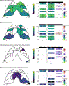
– Statistically significant (FDR adjusted p < .05) changes in z-score distributions for left and right thalamic nuclei and the laterality index in early mild cognitive impairment, late mild cognitive impairment, and Alzheimer’s disease. Values represent the difference in means between the z-score distribution of the control cohort to the phenotypic cohort (see z-score distributions in Figure 3). LI – Laterality Index, calculated as (L-R)/(L+R), where L is the volume of the left region and R is the volume of the right region. A positive value indicates that volumes of the left sided region are smaller than the volumes of the right sided region relative to the control cohort. Thalamus – whole thalamus; AV – anteroventral nucleus; VA – ventral anterior nucleus; VLa – ventral lateral anterior nucleus; VLp – ventral lateral posterior nucleus; VPL – ventral posterior lateral nucleus; Pul – pulvinar nucleus; LGN – lateral geniculate nucleus; MGN – medial geniculate nucleus; CM – centromedian nucleus; MD/Pf – medial dorsal-parafascicular nucleus.
Figure 6
Figure 6
- Statistically significant (FDR adjusted p < .05) changes in z-score distributions for left and right thalamic nuclei and the laterality index in schizophrenia cohorts. Values represent the difference in means between the z-score distribution of the control cohort to the phenotypic cohort (see z-score distributions in Figure 3). LI – Laterality Index, calculated as (L-R)/(L+R), where L is the volume of the left region and R is the volume of the right region. A positive value indicates that volumes of the left sided region are smaller than the volumes of the right sided region relative to the control cohort. Thalamus – whole thalamus; AV – anteroventral nucleus; VA – ventral anterior nucleus; VLa – ventral lateral anterior nucleus; VLp – ventral lateral posterior nucleus; VPL – ventral posterior lateral nucleus; Pul – pulvinar nucleus; LGN – lateral geniculate nucleus; MGN – medial geniculate nucleus; CM – centromedian nucleus; MD/Pf – medial dorsal-parafascicular nucleus.
Figure 7
Figure 7
– Comparison of normative modeling on left side of figure to traditional case-control study on the right side of the figure. Normative modeling values represent the difference in means for statistically significant changes in z-score distributions. Case-control values represent the effect size (Cohen’s d). AV – anteroventral nucleus; VA – ventral anterior nucleus; VLa – ventral lateral anterior nucleus; VLp – ventral lateral posterior nucleus; VPL – ventral posterior lateral nucleus; Pul – pulvinar nucleus; LGN – lateral geniculate nucleus; MGN – medial geniculate nucleus; CM – centromedian nucleus; MD/Pf – medial dorsal-parafascicular nucleus.

Update of

References

    1. Biesbroek JM, Verhagen MG, van der Stigchel S, Biessels GJ (2023): When the central integrator disintegrates: A review of the role of the thalamus in cognition and dementia. Alzheimers Dement. 10.1002/alz.13563 - DOI - PMC - PubMed
    1. Schmahmann JD (2003): Vascular syndromes of the thalamus. Stroke 34: 2264–2278. - PubMed
    1. Neudorfer C, Kultas-Ilinsky K, Ilinsky I, Paschen S, Helmers A-K, Cosgrove GR, et al. (2024): The role of the motor thalamus in deep brain stimulation for essential tremor. Neurotherapeutics 21: e00313. - PMC - PubMed
    1. Fujimori J, Nakashima I (2024): Early-stage volume losses in the corpus callosum and thalamus predict the progression of brain atrophy in patients with multiple sclerosis. J Neuroimmunol 387: 578280. - PubMed
    1. Burdette D, Patra S, Johnson L (2024): Corticothalamic Responsive Neurostimulation for Focal Epilepsy: A Single-Center Experience. J Clin Neurophysiol. 10.1097/WNP.0000000000001060 - DOI - PMC - PubMed