Review
. 2025 Jan 15:24:elae033.
doi: 10.1093/bfgp/elae033.

Environmental community transcriptomics: strategies and struggles

Affiliations
Review

Environmental community transcriptomics: strategies and struggles

Jeanet Mante et al. Brief Funct Genomics. .

Abstract

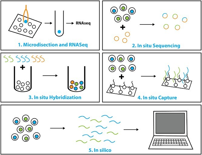
Transcriptomics is the study of RNA transcripts, the portion of the genome that is transcribed, in a specific cell, tissue, or organism. Transcriptomics provides insight into gene expression patterns, regulation, and the underlying mechanisms of cellular processes. Community transcriptomics takes this a step further by studying the RNA transcripts from environmental assemblies of organisms, with the intention of better understanding the interactions between members of the community. Community transcriptomics requires successful extraction of RNA from a diverse set of organisms and subsequent analysis via mapping those reads to a reference genome or de novo assembly of the reads. Both, extraction protocols and the analysis steps can pose hurdles for community transcriptomics. This review covers advances in transcriptomic techniques and assesses the viability of applying them to community transcriptomics.

Keywords: label-free cell sorting; metatranscriptomics; single cell transcriptomics; spatial transcriptomics; transcriptomics.


Conflict of interest statement

The authors declare no conflicts of interest.

Figures

Figure 1
Figure 1
An overview of different community transcriptomics methods. A) Metatranscriptomics: a method for capturing the transcriptomes of all the members of a community at once, B) Spatial Transcriptomics: methods that combine high-resolution imaging with transcriptomics analysis to measure gene expression patterns within a sample while preserving the spatial information of individual cells, C) Sorted Transcriptomics: methods utilize sorting to reduce a complex environmental community into simpler sub-populations that can then be transcriptionally analysed.
Figure 2
Figure 2
General steps for a metatranscriptomic method: 1) Extract RNA from the sample, 2) Enrich mRNA (and synthesize cDNA), 3) sequence the mRNA or cDNA, 4) assemble full transcripts from shorter sequences, 5) assign species information, and 6) predict translation and function of transcripts.
Figure 3
Figure 3
Methods of performing spatial transcriptomics. 1) microdissection followed by RNA-seq. 2)in situ sequencing: rolling circle amplification is used to increase the number of copies and then sequencing is done in situ via fluorescent read out (similar to Sanger sequencing). 3)in situ hybridization: transcripts are identified via fluorescently labelled probes. 4)in situ capture: tissue section is placed on a slide with immobilized primers, the tissue is permeabilized and the transcripts that bind to primers are sequenced. 5)in silico spatial transcriptomics: metatranscriptomics is carried out and computational processing is used to try to reconstruct the spatial dynamics.
Figure 4
Figure 4
This is the sorting of two populations of E. coli measured using a traditional FACS machine (SA3800 Spectral Analyzer). The purple is E. coli W3110, which has a shortened lipopolysacchride (LPS) that makes it slightly smaller than green E. coli ATCC 25922 strain that has a full LPS. It is possible to do gating based on forward and side scatter to sort the two populations. However, a labelled group is required to set the gating parameters, and the overlapping region must be discarded. This data is the unpublished work of the authors.
Figure 5
Figure 5
Different kinds of metabolic functional labelling (before and after digestion). A) Graphene dot that is quenched by the molecule of interest (blue) and fluoresces once the molecule is digested. B) FRET quenching between the molecule of interest (blue) and an appropriate fluorophore (orange). The fluorescence is restored once the molecule of interest is digested. C) FRET between a donor (magenta) and acceptor (orange). One fluorophore is bound to labelling particle via a bond of interest (blue). If the bond of interest is broken the fluorescence stops as the two fluorescent molecules are no longer sufficiently close. D) FRET between a donor (magenta) and acceptor (orange) linked to the molecule of interest (blue). If the molecule of interest is metabolized the donor and acceptor are no longer close enough to maintain FRET.
Figure 6
Figure 6
Ghost Cytometry Method. Panels A and B show the side and front view of the system set up. Note how the laser is split into different frequencies (f1, f2,..fn). These frequencies provide x-axis information while time provides y-axis information. Panel C shows how the waveform obtained can be turned back into a cell image by using a Fourier transform to deconvolute frequencies.

References

    1. Olsen M, Nassar R, Senok A et al. A pilot metagenomic study reveals that community derived mobile phones are reservoirs of viable pathogenic microbes. Sci Rep 2021;11:14102. 10.1038/s41598-021-93622-w. - DOI - PMC - PubMed
    1. Qu Q, Zhang Z, Peijnenburg WJGM et al. Rhizosphere microbiome assembly and its impact on plant growth. J Agric Food Chem 2020;68:5024–38. 10.1021/acs.jafc.0c00073. - DOI - PubMed
    1. Jing H, Xiao X, Zhang Y et al. Composition and ecological roles of the core microbiome along the abyssal–hadal transition zone sediments of the Mariana Trench. Microbiology Spectrum 2022;10:e01988–21. 10.1128/spectrum.01988-21. - DOI - PMC - PubMed
    1. Hurley A, Chevrette MG, Rosario-Meléndez N et al. Thor’s hammer: the antibiotic koreenceine drives gene expression in a model microbial community. MBio 2022;13:e02486–21. 10.1128/mbio.02486-21. - DOI - PMC - PubMed
    1. Raghavan V, Kraft L, Mesny F et al. A simple guide to de novo transcriptome assembly and annotation. Brief Bioinform 2022;23:bbab563. 10.1093/bib/bbab563. - DOI - PMC - PubMed