

Effect of 660-nm LED photobiomodulation on the proliferation and chondrogenesis of meniscus-derived stem cells (MeSCs)
- PMID: 39183213
- PMCID: PMC11345413
- DOI: 10.1038/s41598-024-70258-0
Effect of 660-nm LED photobiomodulation on the proliferation and chondrogenesis of meniscus-derived stem cells (MeSCs)
Abstract
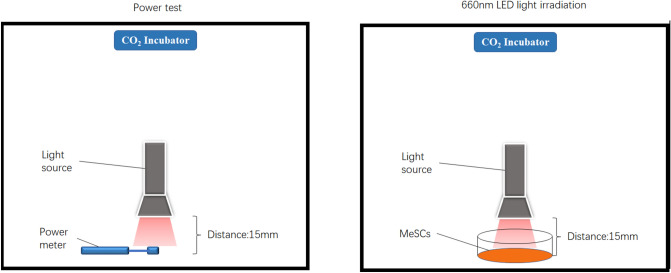
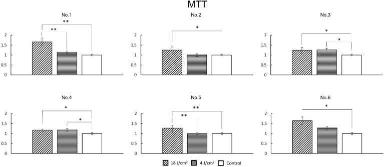
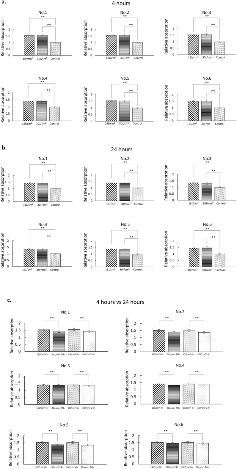
Meniscus-derived stem cells (MeSCs), a unique type of MSC, have outstanding advantages in meniscal cytotherapy and tissue engineering, but the effects and molecular mechanisms of PBM on MeSCs are still unclear. We used 660-nm LED light with different energy densities to irradiate six human MeSC samples and tested their proliferation rate via cell counting, chondrogenic differentiation capacity via the DMMB assay, mitochondrial activity via the MTT assay, and gene expression via qPCR. The proliferation ability, chondrogenic capacity and mitochondrial activity of the 18 J/cm2 group were greater than those of the 4 J/cm2 and control groups. The mRNA expression levels of Akt, PI3K, TGF-β3, Ki67 and Notch-1 in the 18 J/cm2 group were greater than those in the other groups in most samples. After chondrogenic induction, the expression of Col2A1, Sox9 and Aggrecan in the 18 J/cm2 group was significantly greater than that in the 4 J/cm2 and control groups in most of the samples. The variation in the MTT values and Src, PI3K, Akt, mTOR and GSK3β levels decreased with time. The results showed that 660-nm LED red light promoted proliferation and chondrogenic differentiation and affected the gene expression of MeSCs, and the effects on gene expression and mitochondrial activity decreased with time.
© 2024. The Author(s).
Conflict of interest statement
The authors declare no competing interests.
Figures


References
-
- Wiley, T. J. et al. Return to play following meniscal repair. Clin. Sports Med.39, 185–196 (2020). - PubMed
-
- Faucett, S. C. et al. Meniscus root repair vs meniscectomy or nonoperative management to prevent knee osteoarthritis after medial meniscus root tears: Clinical and economic effectiveness. Am. J. Sports Med.47, 762–769 (2019). - PubMed
-
- Suzuki, S. et al. Morphological changes in synovial mesenchymal stem cells during their adhesion to the meniscus. Lab. Invest.100, 916–927 (2020). - PubMed
MeSH terms
Substances
Grants and funding
LinkOut - more resources
Full Text Sources
Research Materials
Miscellaneous
