. 2024 Aug 25;15(1):7323.
doi: 10.1038/s41467-024-51808-6.

Structural basis for allosteric regulation of human phosphofructokinase-1

Affiliations

Structural basis for allosteric regulation of human phosphofructokinase-1

Eric M Lynch et al. Nat Commun. .

Abstract

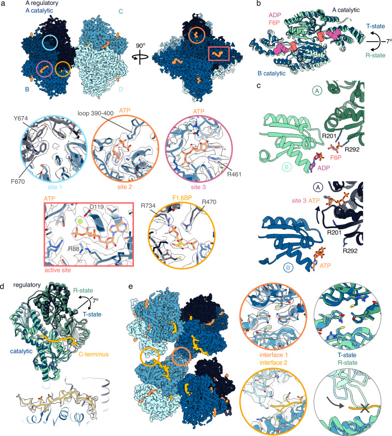
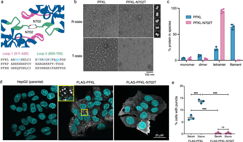
Phosphofructokinase-1 (PFK1) catalyzes the rate-limiting step of glycolysis, committing glucose to conversion into cellular energy. PFK1 is highly regulated to respond to the changing energy needs of the cell. In bacteria, the structural basis of PFK1 regulation is a textbook example of allostery; molecular signals of low and high cellular energy promote transition between an active R-state and inactive T-state conformation, respectively. Little is known, however, about the structural basis for regulation of eukaryotic PFK1. Here, we determine structures of the human liver isoform of PFK1 (PFKL) in the R- and T-state by cryoEM, providing insight into eukaryotic PFK1 allosteric regulatory mechanisms. The T-state structure reveals conformational differences between the bacterial and eukaryotic enzyme, the mechanisms of allosteric inhibition by ATP binding at multiple sites, and an autoinhibitory role of the C-terminus in stabilizing the T-state. We also determine structures of PFKL filaments that define the mechanism of higher-order assembly and demonstrate that these structures are necessary for higher-order assembly of PFKL in cells.


Conflict of interest statement

The authors declare no competing interests.

Figures

Fig. 1
Fig. 1. cryoEM structures of PFKL tetramers and filaments in the R-state.
a Representative 2D class averages of PFKL in the presence of fructose 6-phosphate (F6P) and ATP. b Generalization of the cryoEM processing scheme. Three structures were determined: a free tetramer, a filament, and a consensus tetramer including both free and filament-associated tetramers. c Overlay of the three structures outlined in (b) which are in the same conformation. d CryoEM structure of the consensus PFKL tetramer, colored by monomer. Monomer A is also colored by domain. e Zoomed-in views of the regions indicated in (d) showing the various ligand-binding sites for ADP, F6P, and fructose 1,6-bisphosphate (F1,6BP). f F6P- and ADP-bound PFKL is in the R-state conformation, as revealed by comparison to existing R-state PFK structures. Catalytic domains are shown, with F6P and ADP colored as in panel (e). Structures are aligned on catalytic domain A. g CryoEM structure of the R-state PFKL filament, colored by monomer. Circles show zoomed-in views of filament interfaces 1 and 2. Two views of interface 1 are shown, focused on N702 (left) and F700 (right) at the center of the interface.
Fig. 2
Fig. 2. cryoEM structures of PFKL tetramers and filaments in the T-state.
a CryoEM structure of the consensus PFKL tetramer bound to ATP and fructose 1,6-bisphosphate (F1,6BP), colored by monomer. Monomer A is also colored by domain. Insets show the various ligand binding sites. b Comparison of the catalytic domain conformation of the ATP- and F1,6BP-bound structure (blue) with the R-state PFKL structure (green). Structures are aligned on the catalytic domain of subunit A. ADP and fructose 6-phosphate (F6P) from the R-state structure are shown in the active site. c Comparison of the F6P binding site in the R-state (green) and T-state (blue) structures. The binding site involves residues from the catalytic domains of two monomers (A and B). d Comparison of monomer conformations in the R-state (green) and T-state (blue) structures. Structures are aligned on their catalytic domains (lighter shades). The C-terminus (yellow) bridges across the regulatory and catalytic domains in the T-state conformation. Lower panel shows the isolated density of the C-terminus from the T-state cryoEM map. e CryoEM structure of the T-state PFKL filament, colored by monomer. Circles show zoomed-in views of filament interfaces 1 and 2. Panels at right show comparisons to interfaces 1 and 2 in the R-state structure. Inward rotation of the catalytic domain at interface 2 in the R-state would produce a clash with the C-terminus at the position observed in the T-state structure.
Fig. 3
Fig. 3. Filament interface 1 is essential for assembly of PFKL in vitro and in cells.
a Structure of PFKL filament interface 1 showing two loops forming the interface (top) and sequence alignment of interface 1 residues with other human isoforms (bottom). Poorly-conserved PFKL interface residues are highlighted in blue, with N702 underlined. b Negative stain EM showing that PFKL-N702T does not form filaments in either R-state or T-state ligand conditions. Panels at right show representative 2D class averages of PFKL-N702T tetramers in R-state conditions. Scale bar is 100 nm. c Quantification of assembly state of PFKL and PFKL-N702T by mass photometry. Relative abundance of oligomers was calculated as described in Methods. Data are presented as mean and standard deviation of n = 3 experiments for PFKL and n = 4 experiments for PFKL-N702T, with individual data points also shown. d Immunofluorescent images of parental HepG2 cells and cells expressing wild type FLAG-PFKL or FLAG-PFKL-N702T labeled with anti-FLAG (white) and Hoechst 33342 (blue) under serum starved conditions. Puncta are only visible in wild type FLAG-PFKL cells. Inset shows a higher magnification view of the 10 μm by 10 μm area in the yellow box. Scale bar is 20 µm. e Quantification of the number of cells in continuous serum or in serum starved conditions containing PFKL punctae, as defined as cells with three or greater PFKL assemblies of at least 1 μm diameter. Data is representative of 3 independent passages of cells. Mean and SEM are shown, two-way ANOVA test (PFKL, serum vs starved p = 0.0008; N702T, serum vs staved p = 0.9999; Serum, PFKL vs N702T p = 0.0001; Starved, PFKL vs N702T p = 0.0001).
Fig. 4
Fig. 4. The C-terminal tail of PFKL stabilizes the T-state conformation.
a Enzyme activity assays to determine the affinity for F6P (left) and ATP (right) for wild type PFKL (blue) and PFKL-∆C (green). Assay conditions are described in Table 1. F6P data are mean and SEM of n = 7 replicates for PFKL (n = 6 for 2 mM F6P, n = 1 for 0.25 mM F6P) and n = 5 replicates for PFKL-∆C (n = 2 for 2 mM F6P). ATP data are mean and SEM of n = 7 replicates for PFKL and n = 4 replicates for PFKL-∆C. b Difference in per-residue root-mean-square fluctuations (∆RMSF) after C-terminal tail removal as observed in MD simulations. The light blue regions correspond to the 95% confidence intervals. Arginine residues 88 and 210, involved in binding F6P in the R-state, are highlighted by arrows. c, d PFKL monomer with residues colored according to ∆RMSF values in (b). Removal of the C-terminus produces elevated ∆RMSF values at the C-terminus binding site (c) and in the active site (d). e Average Cα RMSD from the cryoEM structure of a T-state subunit during the last 200 ns of the MD simulations for PFKL and PFKL-∆C. Data points represent values for a total of 12 protein subunits sampled in 3 simulation replicas. Error bars represent the 95% confidence intervals.

Update of

References

    1. Evans, P. R., Farrants, G. W. & Lawrence, M. C. Crystallographic structure of allosterically inhibited phosphofructokinase at 7 A resolution. J. Mol. Biol.191, 713–720 (1986). 10.1016/0022-2836(86)90455-9 - DOI - PubMed
    1. Schirmer, T. & Evans, P. R. Structural basis of the allosteric behaviour of phosphofructokinase. Nature343, 140–145 (1990). 10.1038/343140a0 - DOI - PubMed
    1. Evans, P. R., Farrants, G. W. & Hudson, P. J. Phosphofructokinase: structure and control. Philos. Trans. R. Soc. Lond. B Biol. Sci.293, 53–62 (1981). 10.1098/rstb.1981.0059 - DOI - PubMed
    1. Poorman, R. A., Randolph, A., Kemp, R. G. & Heinrikson, R. L. Evolution of phosphofructokinase—gene duplication and creation of new effector sites. Nature309, 467–469 (1984). 10.1038/309467a0 - DOI - PubMed
    1. Kemp, R. G. & Gunasekera, D. Evolution of the allosteric ligand sites of mammalian phosphofructo-1-kinase. Biochemistry41, 9426–9430 (2002). 10.1021/bi020110d - DOI - PubMed

LinkOut - more resources