. 2024 Jul 17;15(33):13279-13289.
doi: 10.1039/d4sc03299c. eCollection 2024 Aug 22.

Reversible and size-controlled assembly of reflectin proteins using a charged azobenzene photoswitch

Affiliations

Reversible and size-controlled assembly of reflectin proteins using a charged azobenzene photoswitch

Cassidy M Tobin et al. Chem Sci. .

Abstract

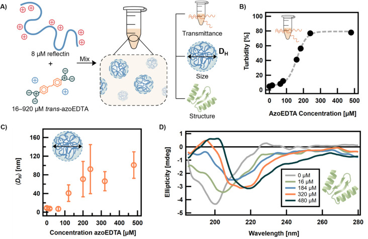
Disordered proteins often undergo a stimuli-responsive, disorder-to-order transition which facilitates dynamic processes that modulate the physiological activities and material properties of cells, such as strength, chemical composition, and reflectance. It remains challenging to gain rapid and spatiotemporal control over such disorder-to-order transitions, which limits the incorporation of these proteins into novel materials. The reflectin protein is a cationic, disordered protein whose assembly is responsible for dynamic color camouflage in cephalopods. Stimuli-responsive control of reflectin's assembly would enable the design of biophotonic materials with tunable color. Herein, a novel, multivalent azobenzene photoswitch is shown to be an effective and non-invasive strategy for co-assembling with reflectin molecules and reversibly controlling assembly size. Photoisomerization between the trans and cis (E and Z) photoisomers promotes or reduces Coulombic interactions, respectively, with reflectin proteins to repeatedly cycle the sizes of the photoswitch-reflectin assemblies between 70 nm and 40 nm. The protein assemblies formed with the trans and cis isomers show differences in interaction stoichiometry and secondary structure, which indicate that photoisomerization modulates the photoswitch-protein interactions to change assembly size. Our results highlight the utility of photoswitchable interactions to control reflectin assembly and provide a tunable synthetic platform that can be adapted to the structure, assembly, and function of other disordered proteins.


Conflict of interest statement

There are no conflicts to declare.

Figures

Fig. 1
Fig. 1. A) Reflectin A1 wildtype protein has alternating, positively charged regions whose Coulombic repulsions initially prevent condensation, folding and formation of higher ordered structures. Charge neutralization overcomes these repulsive interactions, driving associative, non-covalent interactions that form large complexes of reflectin proteins. Counterions on reflectin protein not shown for simplicity. (B) Schematic diagram of photo-controlled electrostatic interactions between reflectin proteins and multivalent azobenzene photoswitches with anionic end groups.
Fig. 2
Fig. 2. (A) Carboxylic acids with varying structure and number of acid groups, but constant valency, were studied for their effects on assembly (counterions not shown for simplicity). The mean hydrodynamic diameters (〈DH〉) of reflectin assemblies prepared as 50 μL samples were measured with DLS. A 20 μM reflectin solution, identically buffered but with no multivalent acid (0), was used as the control. (B) Synthetic scheme for convergent synthesis of azoEDTA with 66% yield. (C) Photoreversible transcis (EZ) isomerization of azoEDTA under weakly acidic conditions. (D) Absorption spectra for 350 μM azoEDTA in 20 mM sodium acetate buffer (pH 4.50) at the photostationary states for 365 nm and 470 nm light.
Fig. 3
Fig. 3. A) Schematic diagram of sample preparation and characterization of assembly of trans-azoEDTA–reflectin protein complexes. Counterions on reflectin protein not shown for simplicity. For a fixed reflectin concentration of 8 μM, the concentration of trans-azoEDTA was varied over the range of 16–920 μM. All samples were prepared in a 20 mM sodium acetate buffer (pH 4.50). (B) Turbidity of the trans-azoEDTA–reflectin system was measured at 400 nm (black dots) and modelled with a sigmoidal function (gray line). (C) The mean hydrodynamic diameter (〈DH〉) of samples measured by DLS. The 〈DH〉 of monomeric, disordered reflectin is 8.9 ± 2.8 nm. Error bars represent standard deviations of average diameter. (D) Ellipticity of azoEDTA–reflectin complexes measured by CD.
Fig. 4
Fig. 4. A) Schematic of sample preparation and characterization of assembly of azoEDTA–reflectin protein complexes for trans-azoEDTA or cis-azoEDTA. For a fixed reflectin concentration of 8 μM, the concentration of trans- and cis-azoEDTA was varied over the range of 80–480 μM. (B) DLS shows change in particle size distribution for azoEDTA–reflectin assemblies with 240 μM azoEDTA. (C) Ratio of moles of azoEDTA to moles of reflectin in the precipitable, assembled protein phase. Error bars represent the standard deviation calculated from 3 independent experiments. (D) Percent of soluble protein remaining in supernatant after centrifugation. Error bars represent the standard deviation calculated from 3 independent experiments.
Fig. 5
Fig. 5. Photoresponse of the azoEDTA–reflectin system for 8 μM reflectin and 240 μM azoEDTA. (A) Schematic illustrating how photoisomerization and turbidity contribute to changes in the measured absorbance of the system upon irradiation. (B) Time-dependent absorbance was collected for 3 cycles with UV light irradiation for 34 min, blue light for 17 min, and no light for 12 min following each irradiation (black dots). The behavior can be fit to a biexponential model (gray lines) that accounts for the photoisomerization and assembly processes. Rate coefficients are given in Table S2. (C) DLS shows a change in assembly size distribution after irradiation with UV and blue light. (D) The change in 〈DH〉, as measured by DLS, can be cycled twice. (E) Evolution of ellipticity of dark-equilibrated azoEDTA–reflectin complexes (I) when exposed to 365 nm (II, IV) and 470 nm (III, V) irradiation. (F) Ellipticity at 202 nm can be cycled as a function of light irradiation.

Similar articles

Cited by

References

    1. Zeng R. Lv C. Wang C. Zhao G. Biotechnol. Adv. 2021;52:107835. doi: 10.1016/j.biotechadv.2021.107835. - DOI - PubMed
    1. Pradhan S. Brooks A. K. Yadavalli V. K. Mater. Today Bio. 2020;7:100065. doi: 10.1016/j.mtbio.2020.100065. - DOI - PMC - PubMed
    1. Chu S. Wang A. L. Bhattacharya A. Montclare J. K. Prog. Biomed. Eng. 2022;4:012003. doi: 10.1088/2516-1091/ac2841. - DOI - PMC - PubMed
    1. Yi D. Bayer T. Badenhorst C. P. S. Wu S. Doerr M. Höhne M. Bornscheuer U. T. Chem. Soc. Rev. 2021;50:8003–8049. doi: 10.1039/D0CS01575J. - DOI - PMC - PubMed
    1. Haynes A. Z. Levine M. Anal. Lett. 2021;54:1871–1880. doi: 10.1080/00032719.2020.1828445. - DOI