. 2024 Jul 22;15(33):13333-13342.
doi: 10.1039/d4sc02703e. eCollection 2024 Aug 22.

Dual inhibitors of Pseudomonas aeruginosa virulence factors LecA and LasB

Affiliations

Dual inhibitors of Pseudomonas aeruginosa virulence factors LecA and LasB

Olga Metelkina et al. Chem Sci. .

Abstract

Dual inhibitors of two key virulence factors of Pseudomonas aeruginosa, the lectin LecA and the protease LasB, open up an opportunity in the current antimicrobial-resistance crisis. A molecular hybridization approach enabled the discovery of potent, selective, and non-toxic thiol-based inhibitors, which simultaneously inhibit these two major extracellular virulence factors and therefore synergistically interfere with virulence. We further demonstrated that the dimerization of these monovalent dual inhibitors under physiological conditions affords divalent inhibitors of LecA with a 200-fold increase in binding affinity. The bifunctional LecA/LasB-blocker 12 showed superiority for the inhibition of virulence mediated by both targets over the individual inhibitors or combinations thereof in vitro. Our study sets the stage for a systematic exploration of dual inhibitors as pathoblockers for a more effective treatment of P. aeruginosa infections and the concept can certainly be extended to other targets and pathogens.


Conflict of interest statement

The authors declare no competing financial interest.

Figures

Fig. 1
Fig. 1. (A) Reported LecA and LasB inhibitors 1–3 and (B) their combination into dual inhibitors.
Fig. 2
Fig. 2. A) Crystal structure of LasB (gray) in complex with 3 (cyan) (PDB code: 7OC7); (B) crystal structure of LecA (petrol) in complex with 1 (magenta) (PDB code: 3ZYF).
Scheme 1
Scheme 1. Synthesis of LecA/LasB dual inhibitors. (a) TfOH, MS 3 Å, DCM, 0 °C, 30 min; (b) H2, Pd/C, DCM, r.t., 18 h; (c) EDCxHCl, DCM, r.t., 4 h; (d) KSAc, acetone, r.t., 2 h; (e) NaOMe, MeOH, r.t., 45 min; (f) NaNO2, 48% HBr, H2O, 0 °C to r.t, 3 h.
Fig. 3
Fig. 3. A) Oxidative dimerization of thiols 11 and 12 into disulfides 26 and 27 using (a) H2O2, DMSO/H2O or (b) P. aeruginosa culture supernatant (yield based on LC-MS); (B) dimerization kinetics of thiols 11 or 12 in the presence of P. aeruginosa culture supernatant into disulfides 26 or 27 analyzed by LC-MS.
Fig. 4
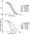
Fig. 4. A) Evaluation of LecA/LasB dual inhibitors by a FRET-based in vitro LasB inhibition assay; (B) evaluation of monomeric LecA/LasB inhibitors 11 and 12 and divalent LecA inhibitors 26a, 26b and 27 and comparison to the control pNP-Gal (1) by a competitive binding assay for LecA based on fluorescence polarization. IC50 values were calculated from at least three independent experiments performed in triplicate.
Fig. 5
Fig. 5. The interaction of dual inhibitors thiol 17 (A) and disulfide 26a (B) with LecA studied using SPR.
Fig. 6
Fig. 6. Crystal structure of 11 in complex with LasB (PDB: 7Z68). (A) Cartoon representation of LasB with a transparent surface (green) and ligated 11 (cyan (R), pink (S)). The amino acids of LasB forming the binding site are represented as sticks. The gray isomesh represents the polder map of 11 contoured at 3σ. (B) Stick representation of the LasB binding site with bound 11. Polar interactions between LasB and 11 are highlighted by dashed lines (color code: oxygen = red, nitrogen = blue, sulfur = yellow, and red spheres = water).
Fig. 7
Fig. 7. Inhibition of the LasB-dependent cytotoxicity of P. aeruginosa PAO1 culture supernatant on human A549 cells. Effect of PAO1 supernatant in the absence (black bar) or presence (grey) of compounds 1, 2, 1 + 2 or 12. The culture supernatant of the lasB knockout mutant (ΔlasB PAO1) was applied as a positive control (light purple). The viability of cells that were untreated (DMEM) served as a negative control and are shown in dark violet. The graphs represent the means of three independent experiments ± SD. One-way ANOVA statistical analysis was performed following Dunnett's multiple comparisons test, comparing the mean value of each concentration to the mean value of PAO1 without any treatment with compounds (***p ≤ 0.001, **p ≤ 0.01, and *p ≤ 0.05).
Fig. 8
Fig. 8. Analysis of the inhibitory activity of 1, 2, a combination of 1 + 2 and 12 on the adhesion of fluorescein-labelled LecA to human A549 cells by fluorescence microscopy. (A) Three representative fluorescence images of one biological replicate of LecA-FITC bound to A549 cells in the presence of the different compounds and under different concentrations (scale bar corresponds to 250 μM). (B) Quantification of mean image fluorescence intensities with the averages and standard deviations for 3 biological replicates ((A), S7A and B†). Intensities are normalized with LecA-FITC in the absence of inhibitors to 100% and in the absence of FITC-LecA to 0%. One-way ANOVA statistical analysis was performed following Dunnett’s multiple comparisons test, comparing the mean value of each condition to the mean value of the LecA-FITC positive control (***p ≤ 0.001, all other data have no statistical significance compared to the LecA-FITC positive control).

References

    1. Miethke M. Pieroni M. Weber T. Brönstrup M. Hammann P. Halby L. Arimondo P. B. Glaser P. Aigle B. Bode H. B. Moreira R. Li Y. Luzhetskyy A. Medema M. H. Pernodet J.-L. Stadler M. Tormo J. R. Genilloud O. Truman A. W. Weissman K. J. Takano E. Sabatini S. Stegmann E. Brötz-Oesterhelt H. Wohlleben W. Seemann M. Empting M. Hirsch A. K. H. Loretz B. Lehr C.-M. Titz A. Herrmann J. Jaeger T. Alt S. Hesterkamp T. Winterhalter M. Schiefer A. Pfarr K. Hoerauf A. Graz H. Graz M. Lindvall M. Ramurthy S. Karlén A. van Dongen M. Petkovic H. Keller A. Peyrane F. Donadio S. Fraisse L. Piddock L. J. V. Gilbert I. H. Moser H. E. Müller R. Nat. Rev. Chem. 2021;5:726–749. doi: 10.1038/s41570-021-00313-1. - DOI - PMC - PubMed
    1. Totsika M. Future Med. Chem. 2017;9:267–269. doi: 10.4155/fmc-2016-0227. - DOI - PubMed
    1. Calvert M. B. Jumde V. R. Titz A. Beilstein J. Org. Chem. 2018;14:2607–2617. doi: 10.3762/bjoc.14.239. - DOI - PMC - PubMed
    1. Clatworthy A. E. Pierson E. Hung D. T. Nat. Chem. Biol. 2007;3:541–548. doi: 10.1038/nchembio.2007.24. - DOI - PubMed
    1. Walesch S. Birkelbach J. Jézéquel G. Haeckl F. P. J. Hegemann J. D. Hesterkamp T. Hirsch A. K. H. Hammann P. Müller R. EMBO Rep. 2023;24:e56033. doi: 10.15252/embr.202256033. - DOI - PMC - PubMed

LinkOut - more resources