

The design of a sample rapid magnetic resonance imaging (MRI) acquisition protocol supporting assessment of multiple articular tissues and pathologies in knee osteoarthritis
- PMID: 39183946
- PMCID: PMC11342198
- DOI: 10.1016/j.ocarto.2024.100505
The design of a sample rapid magnetic resonance imaging (MRI) acquisition protocol supporting assessment of multiple articular tissues and pathologies in knee osteoarthritis
Abstract
Objective: This expert opinion paper proposes a design for a state-of-the-art magnetic resonance image (MRI) acquisition protocol for knee osteoarthritis clinical trials in early and advanced disease. Semi-quantitative and quantitative imaging endpoints are supported, partly amendable to automated analysis. Several (peri-) articular tissues and pathologies are covered, including synovitis.
Method: A PubMed literature search was conducted, with focus on the past 5 years. Further, osteoarthritis imaging experts provided input. Specific MRI sequences, orientations, spatial resolutions and parameter settings were identified to align with study goals. We strived for implementation on standard clinical scanner hardware, with a net acquisition time ≤30 min.
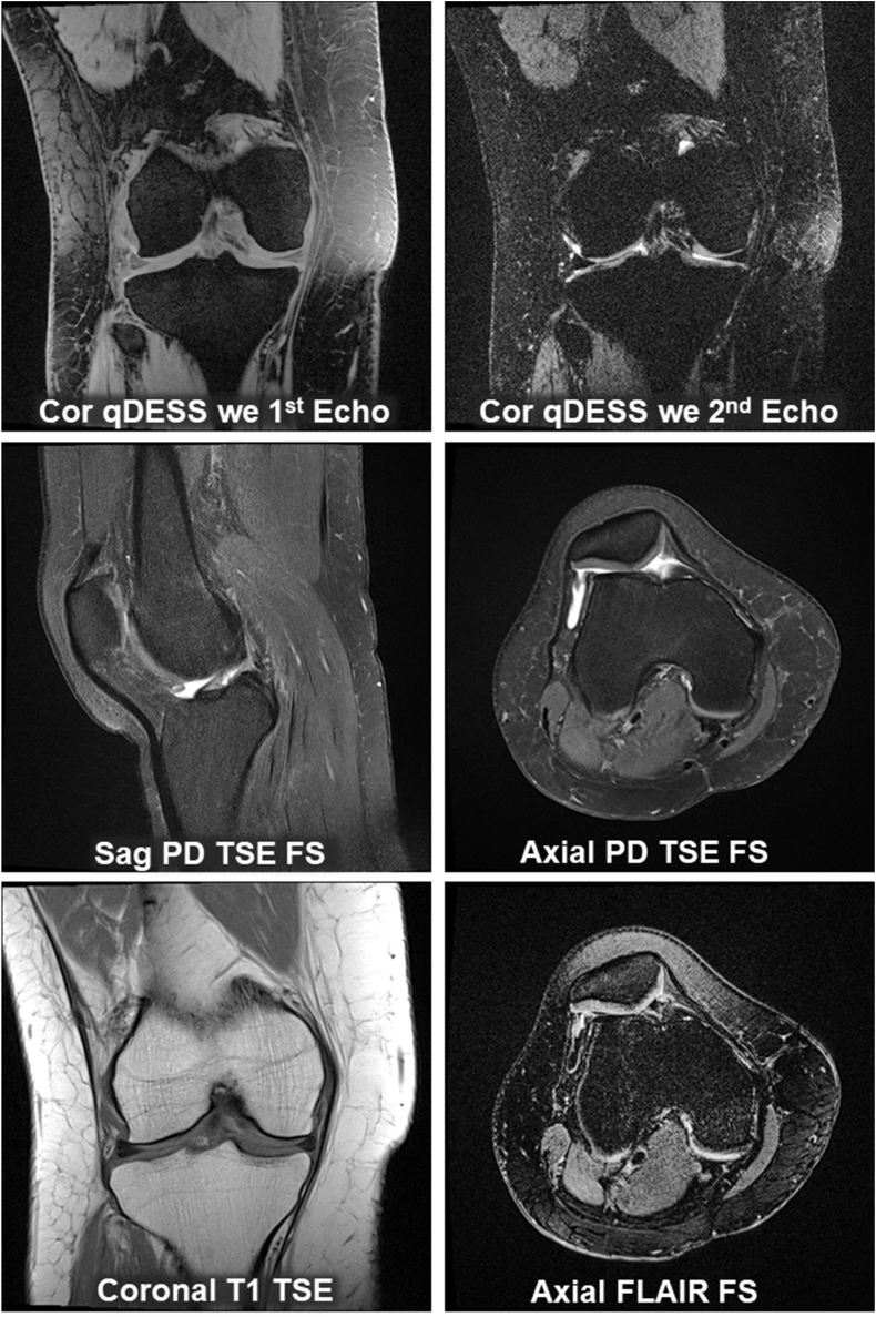
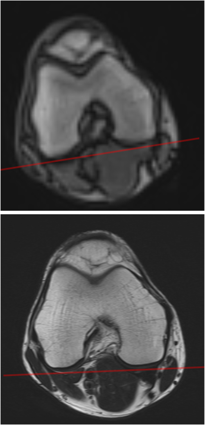
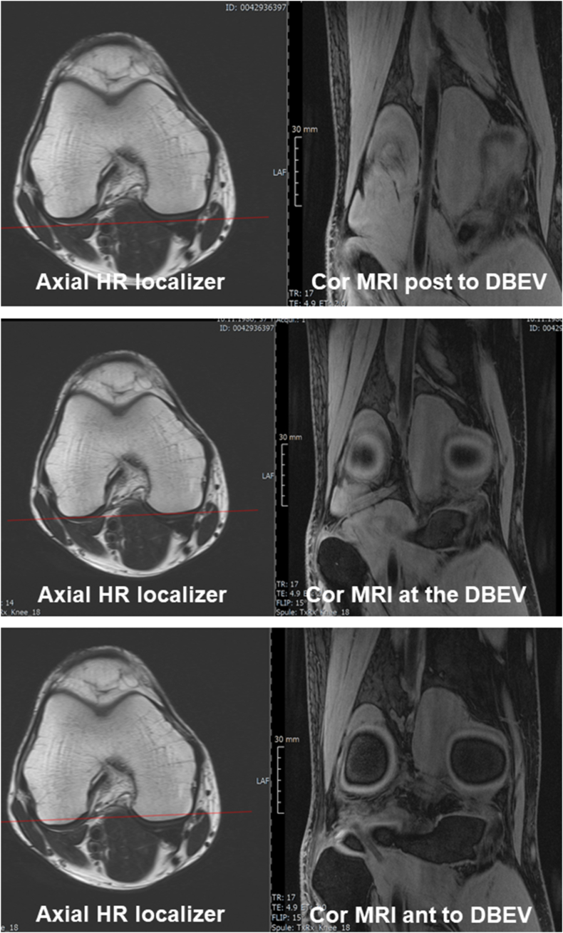
Results: Short- and long-term longitudinal MRIs should be obtained at ≥1.5T, if possible without hardware changes during the study. We suggest a series of gradient- and spin-echo-sequences, supporting MOAKS, quantitative analysis of cartilage morphology and T2, and non-contrast-enhanced depiction of synovitis. These sequences should be properly aligned and positioned using localizer images. One of the sequences may be repeated in each participant (re-test), optimally at baseline and follow-up, to estimate within-study precision. All images should be checked for quality and protocol-adherence as soon as possible after acquisition. Alternative approaches are suggested that expand on the structural endpoints presented.
Conclusions: We aim to bridge the gap between technical MRI acquisition guides and the wealth of imaging literature, proposing a balance between image acquisition efficiency (time), safety, and technical/methodological diversity. This approach may entertain scientific innovation on tissue structure and composition assessment in clinical trials on disease modification of knee osteoarthritis.
Keywords: Clinical trial; Early- and late-stage disease; MRI acquisition protocol; Osteoarthritis (OA); Synovitis.
© 2024 The Author(s).
Conflict of interest statement
FE is CEO/CMO and co-owner of Chondrometrics GmbH; has provided consulting services to Merck KGaA, Kolon-Tissuegene, Servier, Galapagos, Novartis, 4P Pharma/4Moving and Trialspark/Formation Bio; and has – related to this paper – received funding through PROTO from the EU. TCWR has no conflict of interest to declare AC has received research support from the National Institutes of Health, GE Healthcare and Philips and has provided consulting services to Patient Square Capital and Elucid Bioimaging Inc – unrelated to this paper. NMB has – related to this paper – received funding through PROTO from the EU TM has -– related to this paper – received funding through PROTO from the EU GND has – related to this paper – received funding through PROTO from the EU AW is a part-time employee of Chondrometrics GmbH and has – related to this paper – received funding through PROTO from the EU WW is part-time employee and share-holder of Chondrometrics GmbH and has – related to this paper – received funding through PROTO from the EU TW is part of the Executive Board of the Advanced Therapies in Orthopaedics Foundation and has – related to this paper– received funding through PROTO from the EU.
Figures
References
-
- Roemer F.W., Hunter D.J., Crema M.D., Kwoh C.K., Ochoa-Albiztegui E., Guermazi A. An illustrative overview of semi-quantitative MRI scoring of knee osteoarthritis: lessons learned from longitudinal observational studies. Osteoarthritis Cartilage. 2016;24(2):274–289. doi: 10.1016/j.joca.2015.08.011. - DOI - PMC - PubMed
LinkOut - more resources
Full Text Sources
Miscellaneous
