. 2024 Jul 22;27(8):110567.
doi: 10.1016/j.isci.2024.110567. eCollection 2024 Aug 16.

A high-throughput screening identifies MCM chromatin loading inhibitors targeting cells with increased replication origins

Affiliations

A high-throughput screening identifies MCM chromatin loading inhibitors targeting cells with increased replication origins

Lucia Falbo et al. iScience. .

Abstract

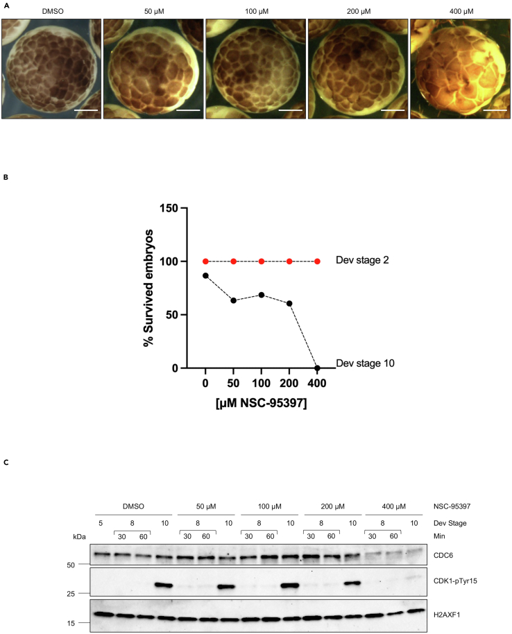
Replication origin assembly is a pivotal step in chromosomal DNA replication. In this process, the ORC complex binds DNA and, together with the CDC6 and CDT1, promotes the loading of the MCM helicase. Chemicals targeting origin assembly might be useful to sensitize highly proliferative cancer cells. However, identifying such compounds is challenging due to the multistage nature of this process. Here, using Xenopus laevis egg extract we set up a high-throughput screening to isolate MCM chromatin loading inhibitors, which led to the identification of NSC-95397 as a powerful inhibitor of replication origin assembly that targets CDC6 protein and promotes its degradation. Using systems developed to test selective drug-induced lethality we show that NSC-95397 triggers cell death both in human cells and Xenopus embryos that have higher proliferative ability. These findings demonstrate the effectiveness of molecules disrupting DNA replication processes in targeting hyperproliferating cells, highlighting their potential as anti-cancer molecules.

Keywords: Biochemistry; Cell biology; Molecular biology.


Conflict of interest statement

The authors declare no competing interests.

Figures

None
Graphical abstract
Figure 1
Figure 1
An in vitro assay to isolate inhibitors of MCM2-7 loading onto DNA (A) Pull-down. Western blot analysis of interphase Xenopus laevis egg extract incubated for 30 min with or without streptavidin beads bound to a 1.8 kb long DNA molecule biotinylated at the 5′-end (bDNA). MCM2-7 binding to DNA was effectively inhibited by Geminin. No DNA indicates the absence of a DNA template, neither sperm nuclei nor the 1.8 kb long DNA fragment. (B) Pull-down of biotinylated DNA (bDNA) was performed at 30 min. HSS was diluted with XB buffer two-to 3-fold in the presence or absence of the recombinant protein Geminin. No DNA indicates the absence of nuclei. (C) Luminescence w/wo geminin. ELISA assay was performed in a 384-well plate using a 2-fold diluted Xenopus extract, in the presence or absence of the recombinant protein Geminin, as well as a crosslinking reagent (formaldehyde). Luminescence was acquired with the Enspire plate reader and it is expressed as Relative Luminescence Unit (RLU). The graph shows a representative experiment.
Figure 2
Figure 2
Setting up a small molecule screening to isolate inhibitors of MCM2-7 loading onto DNA (A) HTS workflow outline. (B) Scatterplot of the primary screening data. The plot of Z-scores for each of the pooled compounds subjected to the primary screen for the identification of an inhibitor of MCM2 loading to bDNA. Values represent a single replicate per compound concentration. The screening was performed on 384-well plates where negative controls (DMSO) are reported in red, positive controls are reported in black (RL5a), and hits are in green. Solid lines represent the mean chemiluminescent value for the measurements of the controls and compounds, and dashed lines denote the average ±3 standard deviation of the negative and positive controls, respectively. (C) Hit confirmation. The pooled compounds were screened in triplicate. Geminin and RL5A were used as positive controls, while DMSO corresponds to the negative control. Data are expressed as the mean percentage of inhibition of MCM7 loading on bDNA compared to vehicle (DMSO). The dashed red line corresponds to the threshold of 50% inhibition. See also Figure S1.
Figure 3
Figure 3
Validation of the identified compounds (A) Chemical structure of the 5 hits identified in the compound screening: NSC-95397, DDP_28574, valsartan, aminacrine, and ethacridine. (B) Western blot analysis of the indicated proteins bound to chromatin isolated from sperm nuclei incubated with DMSO, used as Control, Geminin, or 200 μM NSC-95397, DDP_28574, valsartan, aminacrine, ethacridine for 30 min. (C) Autoradiography of a DNA replication assay showing the time course of α32P-dCTP incorporation in sperm nuclei incubated in egg extract treated with DMSO or with increasing concentrations of NSC-95397 (50-100-150-200 μM). Samples were taken at the indicated times after nuclei and α32P-dCTP addition to egg extracts. Optical density (OD) for each lane is indicated. (D) Representative images of sperm nuclei incubated in interphase extract supplemented with Cy3-dCTP (red) for 30 min (Upper panel) or 90 min (Lower panel) and treated with DMSO, used as control, or 200 μM NSC-95397. Samples were fixed and stained with 4,6-diamidino-2-phenylindole (DAPI) for DNA (blue) and DiOC6 (green) for membranes. Scale bar = 10 μm. See also Figure S2.
Figure 4
Figure 4
A direct effect of NSC-95397 on CDC6 (A) Western blot analysis of the indicated proteins bound to chromatin isolated at the indicated time (1, 5, 15, 30 min) after sperm nuclei addition in egg extract incubated with DMSO, 200 μM NSC-95397, and 200 μM RL5a. No DNA indicates the absence of nuclei. (B) Western blot analysis of the indicated proteins incubated for 5 min at 23°C with 300 μM H2O2, 500 μM roscovitine, 200 μM NSC-95397, or 200 μM MG-132 and then kept on ice. (C) Western blot analysis of the indicated proteins bound to chromatin isolated from sperm nuclei incubated for 30 min with DMSO, used as Control, or 200 μM NSC-95397 analogs (IFM_47059, IFM_47060, IFM_47061, IFM_47062, IFM_47063, IFM_47064, IFM_47065). (D) ITC analysis of NSC-95397 binding to recombinant Xenopus laevis CDC6. See also Figure S3. (E) Representative images of sperm nuclei incubated in interphase extract supplemented with Cy3-dCTP (red) for 90 min with DMSO, used as control, or 100 μM NSC-95397, in the presence of increasing concentrations of recombinant CDC6 protein (0, 80, 160, 320 nM). Samples were fixed and stained with 4,6-diamidino-2-phenylindole (DAPI) for DNA (blue) and DiOC6 (green) for membranes. Scale bar = 10 μm. See also Figure S4. (F) The Cy3-dCTP incorporation of 45 nuclei was quantified as fluorescence-integrated intensity per nucleus using ImageJ. Data are represented as scatter dot plots. Horizontal bars indicate mean ± SEM. A two-way ANOVA test was used to perform multiple comparisons between DMSO and NSC-treated samples with 0 nM of rCDC6 and between NSC-treated samples with 0 nM of rCDC6 and increasing concentrations of recombinant protein added. ∗∗∗∗ = p < 0.0001, ns, not significant.
Figure 5
Figure 5
Effect of NSC-95397 during early development (A) Xenopus eggs were subjected to in vitro fertilization (IVF) and then incubated in the absence (DMSO) or in the presence of NSC-95397 (50 μM, 100 μM, 200 μM or 400 μM), and the survival rate was assessed at 8 h after IVF. Scale bar = 500 μm. (B) The percentage of survived embryos at developmental stage 10 was normalized to the total number of eggs that underwent IVF. (C) Western blot of whole embryos incubated in the absence (DMSO) or in the presence of NSC-95397 (50 μM, 100 μM, 200 μM or 400 μM) and taken at the indicated developmental stages. Stage 8 was sampled every 30 min.
Figure 6
Figure 6
Effects of NSC-95397 in SSRP1 overexpression conditions (A) Images of embryos injected with water (control, first row) or SSRP1 mRNA (second row) and then incubated in the absence (DMSO) or in the presence of 200 μM NSC-95397. Scale bar = 500 μm. See also Figure S5. (B) The percentage of survived embryos at developmental stage 10 was normalized to the total number of eggs that underwent IVF. The results from independent experiments are expressed as the raw percentage of embryos at each developmental stage. Horizontal bars indicate the median with range. One-way ANOVA test was used to analyze differences among multiple groups compared to the control (DMSO). ∗∗ = p < 0.0097, ns = not significant. (C) Western blot of whole embryos injected with water, or Myc-SSRP1 mRNA, treated without (DMSO) or with 200 μM NSC-95397 and taken at the indicated developmental stages.

References

    1. Yuan Z., Schneider S., Dodd T., Riera A., Bai L., Yan C., Magdalou I., Ivanov I., Stillman B., Li H., Speck C. Structural mechanism of helicase loading onto replication origin DNA by ORC-Cdc6. Proc. Natl. Acad. Sci. USA. 2020;117:17747–17756. doi: 10.1073/pnas.2006231117. - DOI - PMC - PubMed
    1. McGarry T.J., Kirschner M.W. Geminin, an inhibitor of DNA replication, is degraded during mitosis. Cell. 1998;93:1043–1053. doi: 10.1016/s0092-8674(00)81209-x. - DOI - PubMed
    1. Li H., O'Donnell M.E. The Eukaryotic CMG Helicase at the Replication Fork: Emerging Architecture Reveals an Unexpected Mechanism. Bioessays. 2018;40 doi: 10.1002/bies.201700208. - DOI - PMC - PubMed
    1. Courtot L., Hoffmann J.S., Bergoglio V. The Protective Role of Dormant Origins in Response to Replicative Stress. Int. J. Mol. Sci. 2018;19 doi: 10.3390/ijms19113569. - DOI - PMC - PubMed
    1. Yekezare M., Gómez-González B., Diffley J.F.X. Controlling DNA replication origins in response to DNA damage - inhibit globally, activate locally. J. Cell Sci. 2013;126:1297–1306. doi: 10.1242/jcs.096701. - DOI - PubMed

LinkOut - more resources