Comparative Evaluation of USG-Guided Single Tissue Marker Versus Multiple Tissue Marker Placements in Breast Malignancy Patients Undergoing Neoadjuvant Chemotherapy for Tumor Localization
- PMID: 39184664
- PMCID: PMC11344559
- DOI: 10.7759/cureus.65355
Comparative Evaluation of USG-Guided Single Tissue Marker Versus Multiple Tissue Marker Placements in Breast Malignancy Patients Undergoing Neoadjuvant Chemotherapy for Tumor Localization
Abstract
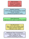
Background Breast cancer remains one of the most common malignancies affecting women globally, contributing significantly to the disease burden. The advent of neoadjuvant chemotherapy (NAC) has revolutionized the treatment for locally advanced breast cancer, allowing tumors to be downstaged and making breast-conserving surgery (BCS) feasible. Accurate localization of the tumor bed post-NAC is crucial for successful surgical removal of residual disease. While traditional single tissue marker placement has been effective, recent advances suggest multiple markers might provide superior localization by comprehensively delineating the entire tumor area. This study aims to compare the effectiveness of single versus multiple tissue marker placements in breast malignancy patients undergoing NAC. Materials and methods A prospective study was conducted in the Department of Radio-diagnosis at Saveetha Medical College over 18 months, including 10 patients diagnosed with breast carcinoma, selected through convenience sampling. Inclusion criteria involved patients diagnosed with breast cancer via mammography, sonography, and histological confirmation, referred for clip placement before NAC. Exclusion criteria were patients unwilling to participate. The procedure involved placing one to two surgical clips within the tumor using a 14/16-gauge coaxial guiding needle under USG guidance, with additional clips for larger or multiple tumors. Data collection included pre-procedural USG, post-procedural mammography (MG1), pre-operative mammography (MG2)/USG, and gross specimen histopathological examination/specimen mammography. Statistical analysis Demographic data, clipping distribution, receptor status, localization methods, surgical outcomes, operation diagnoses, and correlation analysis were statistically analyzed. Mean age, standard deviation, and p-values were calculated to determine the significance of differences between single and multiple clip groups. Results The study included 10 patients with a mean age of 52.5 years. Of these, five (50%) had a single clip, and two (20%) had four clips. The average time from clipping to the second mammogram (MG2) was 106.3 days, and from clipping to operation was 111.0 days, with longer follow-up times for multiple clip patients. Six (60%) of the patients were estrogen receptor (ER) positive, and six (60%) were human epidermal growth factor receptor 2 (HER2) negative. Localization methods were similar between single and multiple clip groups. However, multiple clip patients tended to undergo more extensive surgeries like modified radical mastectomy (MRM). Imaging responses showed no preoperative ultrasound lesions in single clip patients, while multiple clip patients had higher inconsistent diagnoses (10 (100%)) suggesting that multiple clips provide better tumor localization but are linked to increased complexity and longer follow-up times. Conclusion Patients with multiple clips experienced significantly longer follow-up times, reflecting more complex clinical scenarios. Despite no significant differences in receptor status distributions, multiple clip patients required more extensive surgeries, emphasizing the need for tailored surgical planning. The study underscores the importance of considering the number of clips in clinical decision-making. Future research should focus on larger, prospective studies to validate these findings and explore underlying mechanisms.
Keywords: breast carcinoma; clip migration; neoadjuvant chemotherapy; tissue marker; usg-guided procedures.
Copyright © 2024, P et al.
Conflict of interest statement
Human subjects: Consent was obtained or waived by all participants in this study. Saveetha Medical College and Hospital Ethics Committee issued approval ECR/724/Inst/TN/2015/RR-19. Animal subjects: All authors have confirmed that this study did not involve animal subjects or tissue. Conflicts of interest: In compliance with the ICMJE uniform disclosure form, all authors declare the following: Payment/services info: All authors have declared that no financial support was received from any organization for the submitted work. Financial relationships: All authors have declared that they have no financial relationships at present or within the previous three years with any organizations that might have an interest in the submitted work. Other relationships: All authors have declared that there are no other relationships or activities that could appear to have influenced the submitted work.
Figures


References
-
- NCCN guidelines insights: Breast cancer, version 1.2017. Gradishar WJ, Anderson BO, Balassanian R, et al. J Natl Compr Canc Netw. 2017;15:433–451. - PubMed
-
- Role of MRI to assess response to neoadjuvant therapy for breast cancer. Reig B, Heacock L, Lewin A, Cho N, Moy L. J Magn Reson Imaging. 2020;52:249–258. - PubMed
-
- Breast tissue markers: Why? What's out there? How do I choose? Shah AD, Mehta AK, Talati N, Brem R, Margolies LR. Clin Imaging. 2018;52:123–136. - PubMed
LinkOut - more resources
Full Text Sources
Research Materials
Miscellaneous
