[Preprint]. 2025 Mar 6:2023.11.13.566874.
doi: 10.1101/2023.11.13.566874.

Elevated Lactate in the AML Bone Marrow Microenvironment Polarizes Leukemia-Associated Macrophages via GPR81 Signaling

Affiliations

Elevated Lactate in the AML Bone Marrow Microenvironment Polarizes Leukemia-Associated Macrophages via GPR81 Signaling

Celia A Soto et al. bioRxiv. .

Abstract

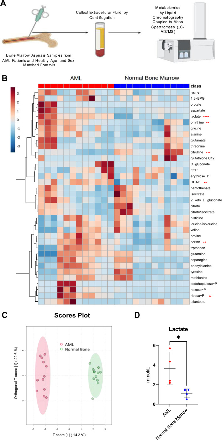
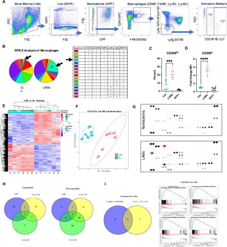
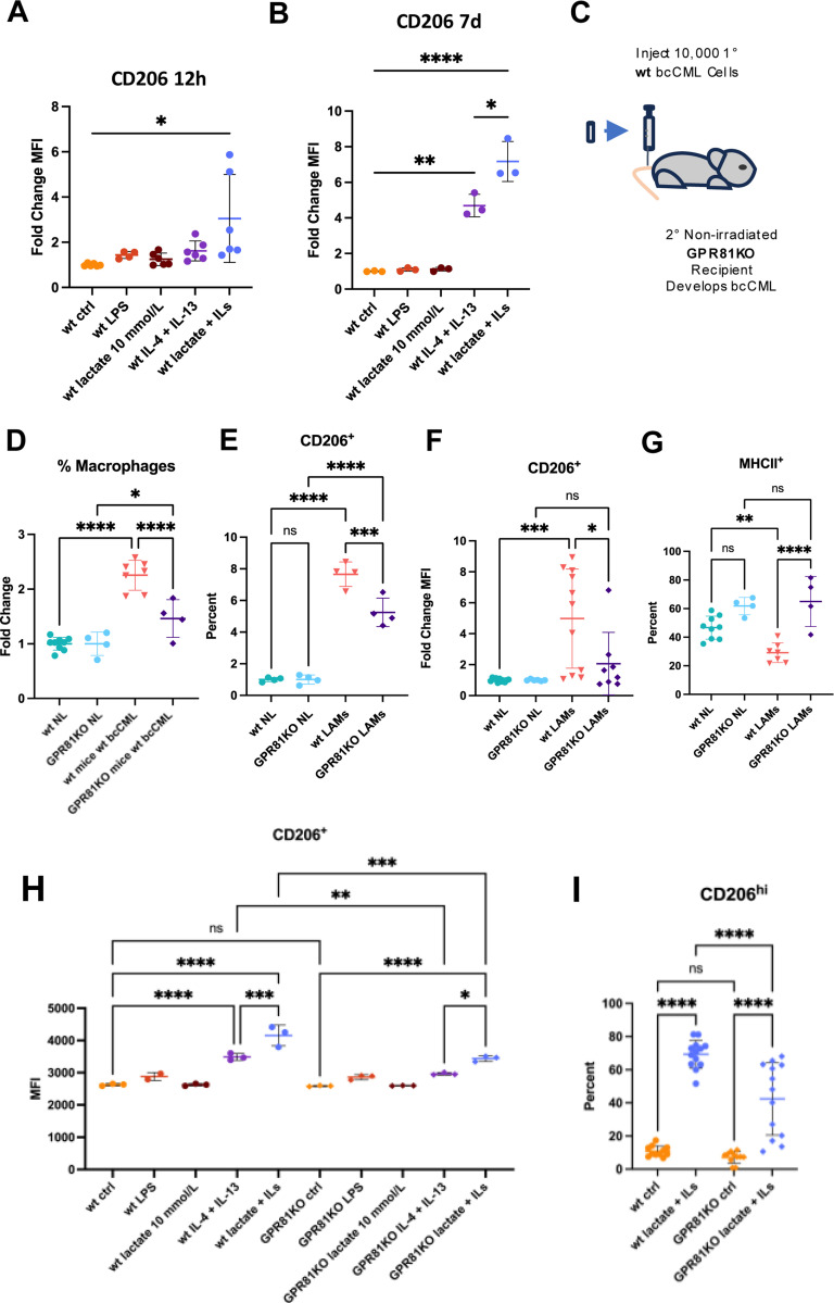
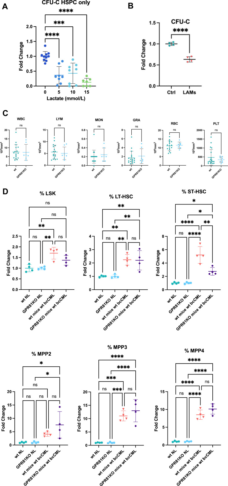
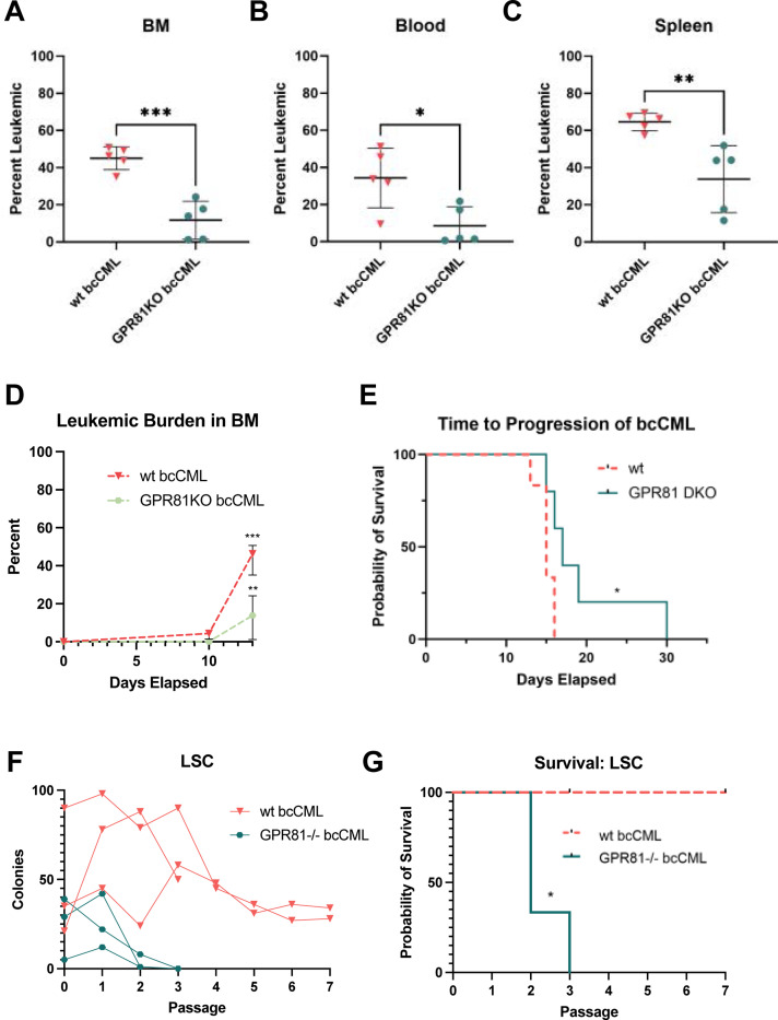
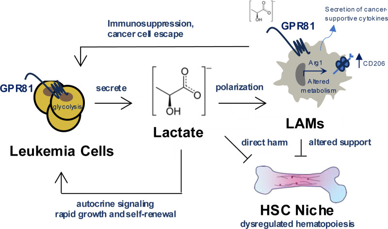
Interactions between acute myeloid leukemia (AML) and the bone marrow microenvironment (BMME) are critical to leukemia progression and chemoresistance. In the solid tumor microenvironment, altered metabolite levels contribute to cancer progression. We performed a metabolomic analysis of AML patient bone marrow serum, revealing increased metabolites compared to age- and sex-matched controls. The most highly elevated metabolite in the AML BMME was lactate. Lactate signaling in solid tumors induces immunosuppressive tumor-associated macrophages and correlates with poor prognosis. This has not yet been studied in the leukemic BMME. Herein, we describe the role of lactate in the polarization of leukemia-associated macrophages (LAMs). Using a murine AML model of blast crisis chronic myelogenous leukemia (bcCML), we characterize the suppressive phenotype of LAMs by surface markers, transcriptomics, and cytokine profiling. Then, mice genetically lacking GPR81, the extracellular lactate receptor, were used to demonstrate GPR81 signaling as a mechanism of both the polarization of LAMs and the direct support of leukemia cells. Furthermore, elevated lactate diminished the function of hematopoietic progenitors and reduced stromal support for normal hematopoiesis. We report microenvironmental lactate as a mechanism of AML-induced immunosuppression and leukemic progression, thus identifying GPR81 signaling as an exciting and novel therapeutic target for treating this devastating disease.


Conflict of interest statement

Conflict of Interest Disclosures Authors have no competing interest to declare.

Figures

Figure 1)
Figure 1). Lactate is elevated in the bone marrow microenvironment (BMME) during acute myeloid leukemia (AML).
A-C, Metabolomics of bone marrow serum from AML patients or normal controls: Graphical depiction of procedure (A), heatmap showing the relative abundance of detectable metabolites (B), and scores plot of principal component analysis (C) (n = 4, in triplicates). D, Lactate concentration in AML and normal bone marrow (BM) serum (n = 4). Significance level determined by unpaired t test for B and D are indicated as: *, P < 0.05; **, P < 0.01; ***, P < 0.001; ****, P < 0.0001. Error bar indicates mean ± standard deviation (SD).
Figure 2)
Figure 2). Leukemia-associated macrophage (LAMs) display an alternatively activated, suppressive phenotype.
A, Gating scheme for flow cytometric analysis of polarization markers on murine macrophages. B, SPICE analysis showing frequency of macrophage subpopulations from healthy controls or late-stage bcCML (n = 5), arrows indicate population enriched in disease. C and D, Nonleukemic control macrophages (Ctrl), leukemia-associated macrophages (LAMs), or leukemia-derived (GFP+) macrophages (n = 7) from bcCML BM. Frequency of CD206hi (C) and expression level by mean fluorescence intensity (MFI) of CD206 on CD206+ (D). E and F, Bulk RNA sequencing of LAMs vs. macrophages from nonleukemic (NL) controls. Heatmap of differentially expressed genes (E) and principal component analysis (PCA) of the top 500 variable genes (F). G, Macrophages were sorted from nonleukemic or leukemic mice and cultured for four days (n = 2), then media was profiled for cytokines. Representative example, arrows indicate a qualitative difference. H, Venn diagrams displaying the number of GO pathways significantly upregulated or downregulated by murine cancer-associated macrophages compared to each study’s own healthy controls: LAMs (n = 6) or tumor-associated macrophages (TAM) from colorectal liver metastasis (CM) (n = 5) or breast cancer (BC) (n = 3). I, Hallmark gene sets enriched in LAMs compared directly to TAMs, determined by gene set enrichment analysis (GSEA): Venn diagram displaying numbers of significantly enriched gene sets, and representative enrichment plots of top significant sets. Significance levels determined by one-way ANOVA for C and D, are indicated as: ***, P < 0.001; ****, P < 0.0001. Error bar indicates mean ± SD. Significance for I was determined by GSEA as an FDR q-value of < 0.25.
Figure 3)
Figure 3). Lactate-GPR81 signaling contributes to leukemia-associated macrophage (LAM) polarization.
A and B, Fold change expression level of CD206 on bone marrow-derived macrophages (BMDMs) in vitro treated with lipopolysaccharide (LPS), lactate, and/or IL-4/−13 (ILs), for for 12 hours (A) (n = 6) or 7 days (B) (n = 3). C, Depiction of the initiation of bcCML in a GPR81KO BMME. D-G, Flow cytometric analysis of nonleukemic (GFP-) bone marrow (BM) cells from bcCML in wt or GPR81KO mice, and nonleukemic (NL) controls. Frequency of macrophages in the BM relative to NL controls of the same genetic background (D) (n = 4–7). BM macrophages: frequency of CD206+ (E), expression level of CD206 relative to NL controls of the same genetic background (F), frequency of MHCII+ (G) (n = 4–11). H, Expression of CD206 on wt or GPR81KO BMDMs treated with LPS, ILs, and/or lactate (n = 3). I, Frequency of CD206hi wt or GPR81KO BMDMs with or without polarization by ILs and lactate. Significance levels determined by one-way ANOVA for A-B, D-G, and H-I are indicated as: ns, not significant; *, P < 0.05; **, P < 0.01; ***, P < 0.001; ****, P < 0.0001. Error bar indicates mean ± SD.
Figure 4)
Figure 4). Excess lactate negatively effects the function of hematopoietic stem and progenitor cells (HSPC) and support, partially regulated by GPR81.
A, HSPCs treated with lactate for 72 hours, fold change colony forming unites (CFU-C) relative to the 0 mmol/L lactate control group (n = 7–10). B, HSPCs cocultured with LAMs or healthy control (Ctrl) macrophages for four days: fold change CFU-Cs relative to control group (n = 4). D, Hematopoietic progenitors’ frequency in the bone marrow, from bcCML in wt or GPR81KO mice, relative to the nonleukemic (NL) control of the same genetic background: hematopoietic stem and progenitor cells (HSPCs/LSK), long-term hematopoietic stem cell (HSC), short-term HSC, multipotent progenitor (MPP) subsets MPP2, MPP3, and MPP4 (n = 4). Significance levels determined by one-way ANOVA for A and D, or by unpaired t test for B are indicated as: ns, not significant; *, P < 0.05; **, P < 0.01; ***, P < 0.001; ****, P < 0.0001. Error bar indicates mean ± SD.
Figure 5)
Figure 5). GPR81 signaling drives leukemic cell expansion rate and leukemia stem cell (LSC) self-renewal.
A-C, Leukemic burden in the bone marrow (BM) (B), peripheral blood (C), and spleen (D), of wt or GPR81KO bcCML, at the timepoint of late-stage disease in wt (n = 5). D, Leukemic burden in the BM over time (n = 5–7). E, Time to progression to late-stage disease in wt or GPR81 DKO bcCML (n = 5). F and G, LSC repopulation, number of colonies at each passage (F), and survival curve (G) (n = 3, in duplicates). Significance levels determined by unpaired t tests for A-D, and log-rank (Mantel-Cox) test for E and G, are indicated as: *, P < 0.05; **, P < 0.01; ***, P < 0.001. Error bar indicates mean ± SD.
Figure 6)
Figure 6). Effects of elevated lactate in the AML bone marrow microenvironment (BMME).
Lactate is elevated in the AML BMME. This polarizes LAMs to a suppressive phenotype, characterized by increased CD206 expression, secretion of cancer-supportive cytokines, expression of Arg1, and altered metabolism. Increased lactate also harms normal hematopoiesis both directly and through altered support by LAMs. Autocrine lactate signaling supports the growth and repopulation of leukemia cells. The lactate receptor GPR81 is a mechanism of LAM polarization, and is implicated in these pathologic mechanisms of AML progression.

References

    1. Ferrara F, Schiffer CA. Acute myeloid leukaemia in adults. The Lancet. 2013;381(9865):484–495. - PubMed
    1. Döhner H, Weisdorf DJ, Bloomfield CD. Acute Myeloid Leukemia. New England Journal of Medicine. 2015;373(12):1136–1152. - PubMed
    1. Chang H-Y, Rodriguez V, Narboni G, Bodey GP, Luna MA, Freireich EJ. CAUSES OF DEATH IN ADULTS WITH ACUTE LEUKEMIA. Medicine. 1976;55(3). - PubMed
    1. Miraki-Moud F, Anjos-Afonso F, Hodby KA, et al. Acute myeloid leukemia does not deplete normal hematopoietic stem cells but induces cytopenias by impeding their differentiation. Proceedings of the National Academy of Sciences of the United States of America. 2013;110(33):13576–13581. - PMC - PubMed
    1. Lapidot T, Sirard C, Vormoor J, et al. A cell initiating human acute myeloid leukaemia after transplantation into SCID mice. Nature. 1994;367(6464):645–648. - PubMed

Publication types