[Preprint]. 2024 Aug 16:2024.08.15.608063.
doi: 10.1101/2024.08.15.608063.

MYH11 rare variant augments aortic growth and induces cardiac hypertrophy and heart failure with pressure overload

Affiliations

MYH11 rare variant augments aortic growth and induces cardiac hypertrophy and heart failure with pressure overload

Zhen Zhou et al. bioRxiv. .

Update in

Abstract

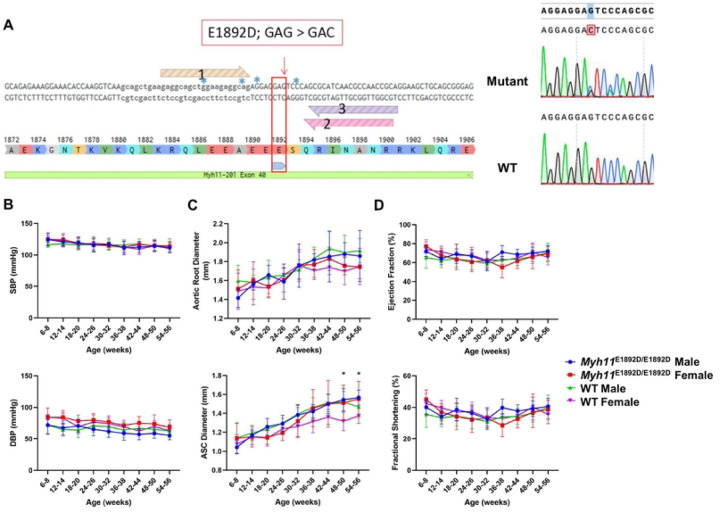
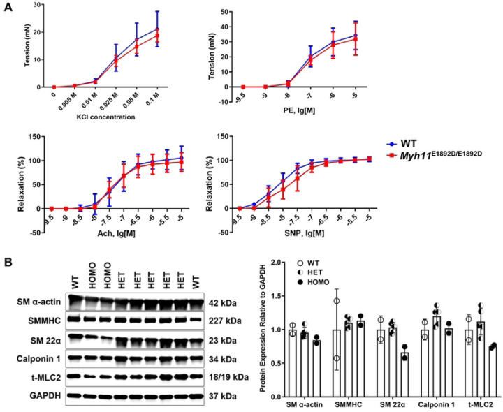
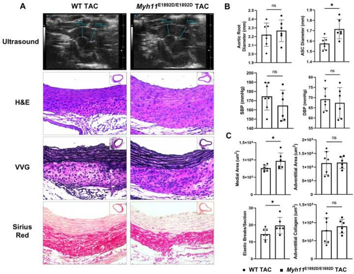
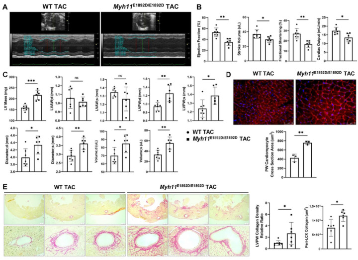
Smooth muscle cell-specific myosin heavy chain, encoded by MYH11, is selectively expressed in smooth muscle cells (SMCs). Pathogenic variants in MYH11 predispose to a number of disorders, including heritable thoracic aortic disease associated with patent ductus arteriosus, visceral myopathy, and megacystis-microcolon-intestinal hypoperistalsis syndrome. Rare variants of uncertain significance occur throughout the gene, including MYH11 p.Glu1892Asp, and we sought to determine if this variant causes thoracic aortic disease in mice. Genomic editing was used to generate Myh11 E1892D/E1892D mice. Wild-type (WT) and mutant mice underwent cardiovascular phenotyping and with transverse aortic constriction (TAC). Myh11 E1892D/E1892D and WT mice displayed similar growth, blood pressure, root and ascending aortic diameters, and cardiac function up to 13 months of age, along with similar contraction and relaxation on myographic testing. TAC induced hypertension similarly in Myh11 E1892D/E1892D and WT mice, but mutant mice showed augmented ascending aortic enlargement and increased elastic fragmentation on histology. Unexpectedly, male Myh11 E1892D/E1892D mice two weeks post-TAC had decreased ejection fraction, stroke volume, fractional shortening, and cardiac output compared to similarly treated male WT mice. Importantly, left ventricular mass increased significantly due to primarily posterior wall thickening, and cardiac histology confirmed cardiomyocyte hypertrophy and increased collagen deposition in the myocardium and surrounding arteries. These results further highlight the clinical heterogeneity associated with MYH11 rare variants. Given that MYH11 is selectively expressed in SMCs, these results implicate a role of vascular SMCs in the heart contributing to cardiac hypertrophy and failure with pressure overload.


Figures

Fig 1.
Fig 1.. Cardiovascular phenotyping in wild-type (WT) and Myh11E1892D/E1892D mice.
(A) Sequencing of DNA sample from tail tissue and cDNA sample from both heart and aorta confirms single nucleotide variant in mouse Myh11 gene. (B) Tail-cuff blood pressure measurement. (C) and (D) Echocardiographic measurements of aortic root, ascending aorta, and left ventricular contractile function. N=10 in each group. SBP, systolic blood pressure, DBP, diastolic blood pressure, ASC, ascending aorta. * P<0.05.
Fig 2.
Fig 2.. Assessment of myograph and smooth muscle cell contractile protein expression in ascending aortic tissues.
(A) Myographic assay of mouse ascending aortic rings at 10 months of age. N=4 (3M+1F) in each group. (B) Immunoblot assay of protein lysates of the ascending aortas from wild-type (WT), heterozygous (HET), and homozygous (HOMO) mice at the age of 6 months. N=2, 5, 2 in the WT, HET, and HOMO group, respectively. KCl, potassium chloride; PE, phenylephrine; Ach, acetylcholine; SNP, sodium nitroprusside.
Fig 3.
Fig 3.. Assessment of ascending aortic remodeling 2 weeks after transverse aortic constriction (TAC) in male mice.
(A) Representative images of proximal aortic ultrasound measurement, H&E, VVG, and Sirius Red staining on ascending aortic tissue sections. (B) TAC induces similar levels of systolic (SBP) and diastolic (DBP) blood pressure in wild-type (WT) and Myh11E1892D/E1892D mice, along with significant ascending aortic enlargement in Myh11E1892D/E1892D mice. (C) Histology analysis shows significantly increased medial thickening and elastic breaks in the mutant aortas compare with WT TAC aortas, with no difference of adventitial area or collagen accumulation. ns, non-significant; * P<0.05. ● WT TAC, ■ Myh11E1892D/E1892D TAC.
Fig 4.
Fig 4.. Assessment of left ventricular remodeling 2 weeks after transverse aortic constriction (TAC) in male mice.
(A) Representative images of left ventricular (LV) contraction using M-mode. (B) Four functional parameters show decreased LV contractility in Myh11E1892D/E1892D mice 2 weeks after TAC. (C) Structural parameters of LV show increased end diastole (d) and systole (s) thickness of posterior wall (PW), along with increased LV diameters and volumes in Myh11E1892D/E1892D mice after TAC. (D) Wheat Germ Agglutinin (WGA) staining of LVPW shows significant increase of cardiomyocyte cross section area in male Myh11E1892D/E1892D heart after TAC. (E) Representative images of Sirius Red staining of LVPW. Quantification of collagen deposition area shows increased peri-arterial (left circumflex artery, LCX) area and LVPW collagen density in Myh11E1892D/E1892D heart after TAC. ns, non-significant; * P<0.05, ** P<0.01, *** P<0.001. ● WT TAC, ■ Myh11E1892D/E1892D TAC.

References

    1. Leguillette R, Lauzon AM. Molecular mechanics of smooth muscle contractile proteins in airway hyperresponsiveness and asthma. Proc Am Thorac Soc. 2008;5(1):40–6. Epub 2007/12/21. doi: 10.1513/pats.200704-053VS. - DOI - PubMed
    1. Miano JM, Cserjesi P, Ligon KL, Periasamy M, Olson EN. Smooth muscle myosin heavy chain exclusively marks the smooth muscle lineage during mouse embryogenesis. Circ Res. 1994;75(5):803–12. Epub 1994/11/01. doi: 10.1161/01.res.75.5.803. - DOI - PubMed
    1. Owens GK. Regulation of differentiation of vascular smooth muscle cells. Physiol Rev. 1995;75(3):487–517. Epub 1995/07/01. doi: 10.1152/physrev.1995.75.3.487. - DOI - PubMed
    1. Manabe I, Owens GK. The smooth muscle myosin heavy chain gene exhibits smooth muscle subtype-selective modular regulation in vivo. J Biol Chem. 2001;276(42):39076–87. Epub 2001/08/08. doi: 10.1074/jbc.M105402200. - DOI - PubMed
    1. Owens GK, Kumar MS, Wamhoff BR. Molecular regulation of vascular smooth muscle cell differentiation in development and disease. Physiol Rev. 2004;84(3):767–801. Epub 2004/07/23. doi: 10.1152/physrev.00041.2003. - DOI - PubMed

Publication types

LinkOut - more resources