

A "glympse" into neurodegeneration: Diffusion MRI and cerebrospinal fluid aquaporin-4 for the assessment of glymphatic system in Alzheimer's disease and other dementias
- PMID: 39185685
- PMCID: PMC11345637
- DOI: 10.1002/hbm.26805
A "glympse" into neurodegeneration: Diffusion MRI and cerebrospinal fluid aquaporin-4 for the assessment of glymphatic system in Alzheimer's disease and other dementias
Erratum in
-
Correction to "A 'Glympse' Into Neurodegeneration: Diffusion MRI and Cerebrospinal Fluid Aquaporin-4 for the Assessment of Glymphatic System in Alzheimer's Disease and Other Dementias".Hum Brain Mapp. 2024 Oct;45(14):e70036. doi: 10.1002/hbm.70036. Hum Brain Mapp. 2024. PMID: 39351889 Free PMC article. No abstract available.
Abstract
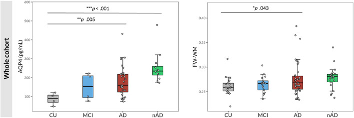
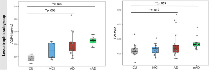
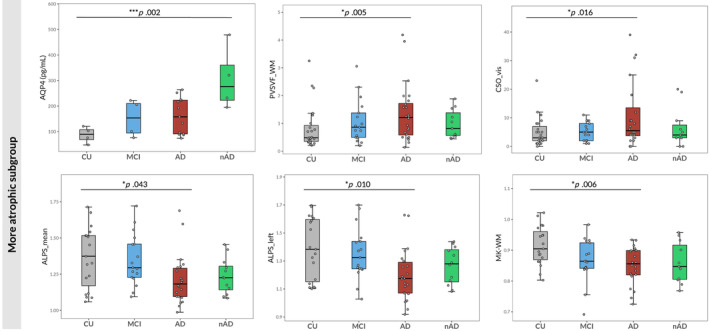
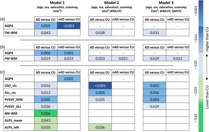
The glymphatic system (GS) is a whole-brain perivascular network, consisting of three compartments: the periarterial and perivenous spaces and the interposed brain parenchyma. GS dysfunction has been implicated in neurodegenerative diseases, particularly Alzheimer's disease (AD). So far, comprehensive research on GS in humans has been limited by the absence of easily accessible biomarkers. Recently, promising non-invasive methods based on magnetic resonance imaging (MRI) along with aquaporin-4 (AQP4) quantification in the cerebrospinal fluid (CSF) were introduced for an indirect assessment of each of the three GS compartments. We recruited 111 consecutive subjects presenting with symptoms suggestive of degenerative cognitive decline, who underwent 3 T MRI scanning including multi-shell diffusion-weighted images. Forty nine out of 111 also underwent CSF examination with quantification of CSF-AQP4. CSF-AQP4 levels and MRI measures-including perivascular spaces (PVS) counts and volume fraction (PVSVF), white matter free water fraction (FW-WM) and mean kurtosis (MK-WM), diffusion tensor imaging analysis along the perivascular spaces (DTI-ALPS) (mean, left and right)-were compared among patients with AD (n = 47) and other neurodegenerative diseases (nAD = 24), patients with stable mild cognitive impairment (MCI = 17) and cognitively unimpaired (CU = 23) elderly people. Two runs of analysis were conducted, the first including all patients; the second after dividing both nAD and AD patients into two subgroups based on gray matter atrophy as a proxy of disease stage. Age, sex, years of education, and scanning time were included as confounding factors in the analyses. Considering the whole cohort, patients with AD showed significantly higher levels of CSF-AQP4 (exp(b) = 2.05, p = .005) and FW-WM FW-WM (exp(b) = 1.06, p = .043) than CU. AQP4 levels were also significantly higher in nAD in respect to CU (exp(b) = 2.98, p < .001). CSF-AQP4 and FW-WM were significantly higher in both less atrophic AD (exp(b) = 2.20, p = .006; exp(b) = 1.08, p = .019, respectively) and nAD patients (exp(b) = 2.66, p = .002; exp(b) = 1.10, p = .019, respectively) compared to CU subjects. Higher total (exp(b) = 1.59, p = .013) and centrum semiovale PVS counts (exp(b) = 1.89, p = .016), total (exp(b) = 1.50, p = .036) and WM PVSVF (exp(b) = 1.89, p = .005) together with lower MK-WM (exp(b) = 0.94, p = .006), mean and left ALPS (exp(b) = 0.91, p = .043; exp(b) = 0.88, p = .010 respectively) were observed in more atrophic AD patients in respect to CU. In addition, more atrophic nAD patients exhibited higher levels of AQP4 (exp(b) = 3.39, p = .002) than CU. Our results indicate significant changes in putative MRI biomarkers of GS and CSF-AQP4 levels in AD and in other neurodegenerative dementias, suggesting a close interaction between glymphatic dysfunction and neurodegeneration, particularly in the case of AD. However, the usefulness of some of these biomarkers as indirect and standalone indices of glymphatic activity may be hindered by their dependence on disease stage and structural brain damage.
Keywords: Alzheimer disease; aquaporin 4; brain perivascular spaces; cerebrospinal fluid; dementia; diffusion magnetic resonance imaging; glymphatic system; neurodegenerative disease.
© 2024 The Author(s). Human Brain Mapping published by Wiley Periodicals LLC.
Conflict of interest statement
The authors declare that they have no competing interests.
Figures
References
-
- Alzheimer's disease Neuroimaging Initiative1 , Zhang, N. , Song, X. , Zhang, Y. , Chen, W. , D'Arcy, R. C. N. , Darvesh, S. , Fisk, J. D. , & Rockwood, K. (2011). An MRI brain atrophy and lesion index to assess the progression of structural changes in Alzheimer's disease, mild cognitive impairment, and Normal aging: A follow‐up study. Journal of Alzheimer's Disease, 26, 359–367. - PubMed
-
- Arighi, A. , Arcaro, M. , Fumagalli, G. G. , Carandini, T. , Pietroboni, A. M. , Sacchi, L. , Fenoglio, C. , Serpente, M. , Sorrentino, F. , Isgrò, G. , Turkheimer, F. , Scarpini, E. , & Galimberti, D. (2022). Aquaporin‐4 cerebrospinal fluid levels are higher in neurodegenerative dementia: Looking at glymphatic system dysregulation. Alzheimer's Research & Therapy, 14, 135. - PMC - PubMed
-
- Armstrong, M. J. , Litvan, I. , Lang, A. E. , Bak, T. H. , Bhatia, K. P. , Borroni, B. , Boxer, A. L. , Dickson, D. W. , Grossman, M. , Hallett, M. , Josephs, K. A. , Kertesz, A. , Lee, S. E. , Miller, B. L. , Reich, S. G. , Riley, D. E. , Tolosa, E. , Tröster, A. I. , Vidailhet, M. , & Weiner, W. J. (2013). Criteria for the diagnosis of corticobasal degeneration. Neurology, 80, 496–503. - PMC - PubMed
MeSH terms
Substances
Grants and funding
LinkOut - more resources
Full Text Sources
Medical
Research Materials
