Review
. 2024 Sep 1;72(9):1244-1253.
doi: 10.4103/IJO.IJO_3236_23. Epub 2024 Aug 23.

Glaucoma and refractive surgery: A comprehensive review

Affiliations
Review

Glaucoma and refractive surgery: A comprehensive review

Sivani Kodali et al. Indian J Ophthalmol. .

Abstract

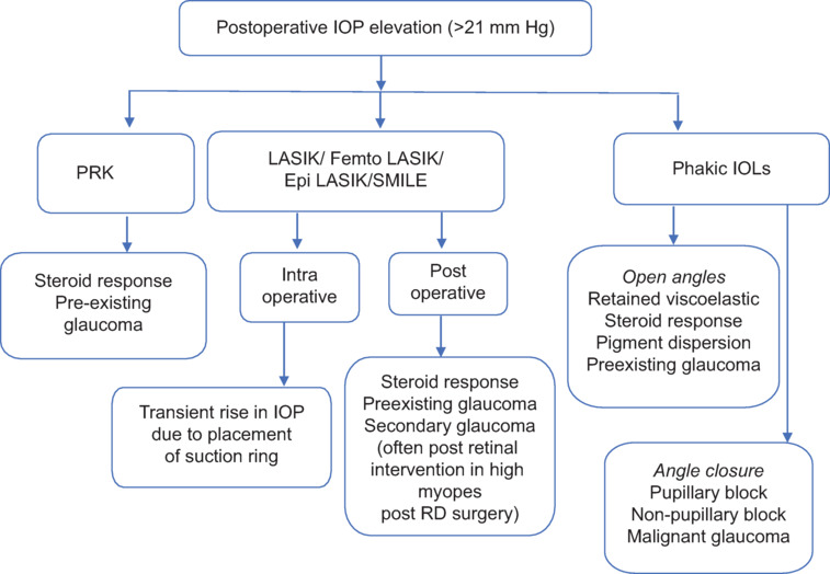
The global surge in refractive surgeries, particularly among myopic individuals, has elicited concerns regarding potential inaccuracies in the measurement of intraocular pressure (IOP) post surgery, primarily associated with central corneal thinning. The incidence of elevated IOP after intraocular refractive surgeries is higher than expected and is multifactorial. Myopic eyes have a higher susceptibility to both primary and secondary glaucoma. Consequently, meticulous preoperative screening for glaucoma is imperative, coupled with systematic postoperative follow-up and evaluation. This comprehensive review analyses the etiology, mechanisms, and therapeutic strategies for managing elevated IOP following refractive surgery. We propose an algorithm to summarize the causative factors of elevated IOP and formulate effective interventions in these instances.


Conflict of interest statement

There are no conflicts of interest.

Figures

Figure 1
Figure 1
Summary of various causes for IOP elevation post refractive surgery are outlined in the flowchart
Figure 2
Figure 2
(a) Clinical image showing grade two subepithelial corneal haze 3 months post photorefractive kearatotomy (PRK). (b) AS-OCT showing hyperreflectivity in the anterior stroma with relatively clear posterior stroma
Figure 3
Figure 3
Slit-lamp photograph showing diffuse lamellar keratitis post small-incision lenticule extraction (SMILE). Diffuse cellular infiltration in the interface was noted in the interface extending to the edge of the flap, which was treated with frequent topical steroids
Figure 4
Figure 4
A 22-year-old, myope, post LASIK has interface fluid syndrome (IFS). (a) Slit view of the cornea showing fluid in the interface. IOP measured in the central cornea with Goldmann applanation tonometry was 18 mmHg, while it was 28 mmHg in the periphery (outside the flap). (b) AS-OCT showing the accumulation of interface fluid in the flap-bed interface (c) Slit view of the cornea with resolved interface fluid with control of intraocular pressure with glaucoma medications. (d) The AS-OCT showing resolved interface fluid
Figure 5
Figure 5
The correct orientation of ICL with a flat surface of the central vault (solid arrow) and posterior convex surface (arrowhead)
Figure 6
Figure 6
(a) Slit-lamp photograph on retroillumination showing a well-placed ICL with two peripheral iridotomies. (b) Very shallow anterior chamber due to pupillary block due to incomplete peripheral iridotomy in an eye with V4b model ICL. (c) Note the shallow anterior chamber and excess ICL vault on AS-OCT
Figure 7
Figure 7
Showing the high vault of ICL of 1456 microns
Figure 8
Figure 8
Shows increased pigment dispersion over the ICL (Asterix) with patent contraflow opening (a and b), note a gap between the ICL and the iris with a shadow (arrow). UBM c shows the reversed orientation of the ICL with a flat surface of the central lens posteriorly and the anterior concave surface. d Note the peripheral tenting of the iris caused by the reversed ICL (arrow)
Figure 9
Figure 9
The management algorithm for post-ICL elevated IOP is shown in a flowchart
Figure 10
Figure 10
A 25-year-old, high myope, post implantable collamer lens (ICL) presented with uncontrolled IOP, diagnosed to have possible preexisting JOAG. (a and b) Large disc with 0.9 CDR with bipolar thin rims with near-notch and diffuse NFL loss as seen on the red-free photo. (c) The visual field shows incomplete biarcuate scotoma, Inferior > superior. (d-f) Underwent trabeculectomy with Ologen implantation. There was good bleb, deep anterior chamber, and stable ICL. The IOP was 15 mmHg with nil medication 3 years post surgery and stable visual field

References

    1. Singh G. Refractive corrective surgery for highly myopic eyes. Ann Transl Med. 2022;10:759. - PMC - PubMed
    1. Kersey JP, Broadway DC. Corticosteroid-induced glaucoma: A review of the literature. Eye (Lond) 2006;20:407–16. - PubMed
    1. Mitchell P, Hourihan F, Sandbach J, Wang JJ. The relationship between glaucoma and myopia: The Blue Mountains eye study. Ophthalmology. 1999;106:2010–5. - PubMed
    1. Xu L, Wang Y, Wang S, Wang Y, Jonas JB. High myopia and glaucoma susceptibility the Beijing Eye Study. Ophthalmology. 2007;114:216–20. - PubMed
    1. Perkins ES, Phelps CD. Open angle glaucoma, ocular hypertension, low-tension glaucoma, and refraction. Arch Ophthalmol. 1982;100:1464–7. - PubMed