. 2024 Aug 1;65(10):34.
doi: 10.1167/iovs.65.10.34.

Agonism of β3-Adrenoceptors Inhibits Pathological Retinal Angiogenesis in the Model of Oxygen-Induced Retinopathy

Affiliations

Agonism of β3-Adrenoceptors Inhibits Pathological Retinal Angiogenesis in the Model of Oxygen-Induced Retinopathy

Alberto Melecchi et al. Invest Ophthalmol Vis Sci. .

Retraction in

Abstract

Purpose: In response to hypoxia, sympathetic fibers to the retina activate β-adrenoceptors (β-ARs) that play an important role in the regulation of vascular and neuronal functions. We investigated the role of β3-AR using the mouse model of oxygen-induced retinopathy (OIR).

Methods: Mouse pups were exposed to 75% oxygen at postnatal day 7 (PD7) followed by a return to room air at PD12. The β3-AR preferential agonist BRL37344 was subcutaneously administered once daily at different times after the return to room air. At PD17, the OIR mice underwent flash and pattern electroretinogram. After sacrifice, retinal wholemounts were used for vessel staining or immunohistochemistry for astrocytes, Müller cells, or retinal ganglion cells (RGCs). In retinal homogenates, the levels of markers associated with neovascularization (NV), the blood-retinal barrier (BRB), or astrocytes were determined by western blot, and quantitative reverse-transcription polymerase chain reaction was used to assess β3-AR messenger. Administration of the β3-AR antagonist SR59230A was performed to verify BRL37344 selectivity.

Results: β3-AR expression is upregulated in response to hypoxia, but its increase is prevented by BRL37344, which counteracts NV by inhibiting the pro-angiogenic pathway, activating the anti-angiogenic pathway, recovering BRB-associated markers, triggering nitric oxide production, and favoring revascularization of the central retina through recovered density of astrocytes that ultimately counteracts NV in the midperiphery. Vasculature rescue prevents dysfunctional retinal activity and counteracts OIR-associated retinal ganglion cell loss.

Conclusions: β3-AR has emerged as a crucial intermediary in hypoxia-dependent NV, suggesting a role of β3-AR agonists in the treatment of proliferative retinopathies.


Conflict of interest statement

Disclosure: A. Melecchi, None; A. Canovai, None; R. Amato, None; M. Dal Monte, None; L. Filippi, None; P. Bagnoli, None; M. Cammalleri, None

Figures

Figure 1.
Figure 1.
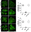
BRL37344 promotes central retina revascularization and reduces the neovascular tuft area. (AD) Representative images of isolectin B4–labeled superficial plexuses of retinas from mice that were normoxic, OIR untreated, or treated with BRL37344 either alone or in combination with the β3-AR antagonist SR59230A. Scale bar: 1 mm. (ad) High magnification of the boxed areas in A to D. Scale bar: 500 µm. The extent of both the avascular area (D) and the tuft area (E) was quantitatively evaluated in OIR mice untreated or treated with BRL37344 either alone or in combination with SR59230A. Data are shown as box plots with minimum to maximum whiskers (n = 6 retinas). *P < 0.05 versus normoxic.
Figure 2.
Figure 2.
BRL37344 affects hypoxia-driven angiogenic markers. (A) Representative western blots and (BG) relative densitometric analysis of HIF-1α, VEGF, PEDF, nNOS, eNOS, and iNOS protein levels in retinal homogenates from normoxic and OIR mice untreated or treated with BRL37344. β-Actin was used as the loading control. Data are shown as box plots with minimum to maximum whiskers (n = 6 samples). *P < 0.05 versus normoxic; §P< 0.05 versus OIR.
Figure 3.
Figure 3.
BRL37344 efficacy on hypoxia-driven angiogenic markers persists up to PD21. (A) Representative western blots and (B, C) relative densitometric analysis of HIF-1α and vascular endothelial growth factor A (VEGFA) protein levels in retinal homogenates from normoxic and OIR mice untreated or treated with BRL37344 from PD12 to PD16. Measurements were performed at PD17, PD21, and PD25. β-Actin was used as the loading control. Data are shown as box plots with minimum to maximum whiskers (n = 6 samples). *P < 0.05 versus normoxic; §P < 0.05 versus OIR.
Figure 4.
Figure 4.
BRL37344 prevents OIR-associated loss of tight-junction proteins. Representative western blots (A) and relative densitometric analysis of ZO-1 (B) and claudin-5 (C) protein levels in retinal homogenates from normoxic and OIR mice untreated or treated with BRL37344. β-Actin was used as the loading control. Data are shown as box plots with minimum to maximum whiskers (n = 6 samples). *P < 0.05 versus normoxic; §P < 0.05 versus OIR.
Figure 5.
Figure 5.
BRL37344 prevents ERG dysfunction. (A) Representative ERG waveforms recorded at a light intensity of 1 log cd·s/m2 in normoxic and OIR mice untreated or treated with BRL37344. (B, C) Scotopic a-wave (B) and b-wave (C) amplitudes plotted as a function of increasing light intensity in normoxic mice (red circles and red line) and OIR mice untreated (black circles and black line) or treated with subcutaneous BRL37344 (green squares and green line). In untreated OIR mice, the amplitudes of the a- and b-waves were lower than those in the normoxic controls. Each point represents the mean ± SEM of data from six mice. *P < 0.05 versus normoxic; §P < 0.05 versus OIR.
Figure 6.
Figure 6.
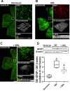
BRL37344 recovers astrocytes density in the central retina and attenuates OIR-induced gliosis. (AC) Representative images of wholemount retinas stained with isolectin B4 from normoxic and OIR mice untreated or treated (left panel). Scale bar: 1 mm. Boxed areas correspond to high-magnification images of the central retina showing GFAP-IR (astrocytes in green, Müller cells in red). Scale bars: 50 µm. The 3D reconstructions and Z-stack projections of GFAP-IR display astrocyte profiles and reactive Müller cells processes, respectively. Scale bars: 50 µm (3D reconstructions) and 70 µm (Z-stack projections). (D) Representative western blots and relative densitometric analysis of protein levels of GFAP in retinal homogenates from normoxic and OIR mice untreated or treated with BRL37344. β-Actin was used as the loading control. Data are shown as box plots with minimum to maximum whiskers (n = 6 samples). *P < 0.05 versus normoxic; §P < 0.05 versus OIR.
Figure 7.
Figure 7.
BRL37344 increases the levels of astrocyte-associated transcription factors. (A, B) Representative western blots and relative densitometric analyses of HIF-2α (A) and PAX2 (B) in retinal homogenates from normoxic and OIR mice untreated or treated with BRL37344. β-actin was used as loading control. Data are shown as box plots with minimum to maximum whiskers (n = 6 samples). *P < 0.05 versus normoxic; §P < 0.05 versus OIR.
Figure 8.
Figure 8.
BRL37344 attenuates OIR-associated RGC loss. (A) Representative images of RBPMS-labeled RGCs in wholemount retinas from normoxic and OIR untreated or treated mice (left panel). Scale bar: 1 mm. Boxed areas correspond to high-magnification images of the central retina displaying RBPMS labeling counterstained with isolectin B4 (right panel). Scale bar: 25 µm. (B) Quantitative evaluation of RBPMS-labeled cell density. Data are shown as box plots with minimum to maximum whiskers (n = 6 retinas). *P < 0.05 versus normoxic; §P < 0.05 versus OIR.
Figure 9.
Figure 9.
BRL37344 prevents OIR-associated PERG alteration. (A) Representative PERG waveforms in normoxic and OIR mice untreated or treated with BRL37344. (B, C) Quantitative analysis of N1–P1 and P1–N2 amplitudes. Data are shown as box plots with minimum to maximum whiskers (n = 6 mice). *P < 0.05 versus normoxic; §P < 0.05 versus OIR.
Figure 10.
Figure 10.
BRL37344 restrains the upregulation of HIF-1α and β3-AR over the hypoxic phase. (A, C) Representative western blots and relative densitometric analysis of HIF-1α and β3-AR protein levels in normoxic and OIR mice untreated or treated with BRL37344. β-Actin was used as the loading control. (B) Levels of β3-AR mRNA as determined by qPCR. Data are shown as box plots with minimum to maximum whiskers (n = 6 samples). *P < 0.05 versus normoxic; §P < 0.05 versus OIR.

References

    1. Shi S, Ding C, Zhu S, et al.. PERK inhibition suppresses neovascularization and protects neurons during ischemia-induced retinopathy. Invest Ophthalmol Vis Sci . 2023; 64(11): 17. - PMC - PubMed
    1. Slavi N, Toychiev AH, Kosmidis S, et al.. Suppression of connexin 43 phosphorylation promotes astrocyte survival and vascular regeneration in proliferative retinopathy. Proc Natl Acad Sci USA . 2018; 115(26): E5934–E5943. - PMC - PubMed
    1. Fu Z, Meng SS, Burnim SB, Smith LE, Lo AC.. Lutein facilitates physiological revascularization in a mouse model of retinopathy of prematurity. Clin Exp Ophthalmol . 2017; 45(5): 529–538. - PMC - PubMed
    1. Dorrell MI, Aguilar E, Jacobson R, et al.. Maintaining retinal astrocytes normalizes revascularization and prevents vascular pathology associated with oxygen-induced retinopathy. Glia. 2010; 58(1): 43–54. - PMC - PubMed
    1. Casini G, Dal Monte M, Fornaciari I, Filippi L, Bagnoli P.. The β-adrenergic system as a possible new target for pharmacologic treatment of neovascular retinal diseases. Prog Retin Eye Res . 2014; 42: 103–129. - PubMed

Publication types

MeSH terms