. 2024 Sep 11;146(36):25232-25244.
doi: 10.1021/jacs.4c09267. Epub 2024 Aug 26.

Bimolecular Sandwich Aggregates of Porphyrin Nanorings

Affiliations

Bimolecular Sandwich Aggregates of Porphyrin Nanorings

Henrik Gotfredsen et al. J Am Chem Soc. .

Abstract

Extended π-systems often form supramolecular aggregates, drastically changing their optical and electronic properties. However, aggregation processes can be difficult to characterize or predict. Here, we show that butadiyne-linked 8- and 12-porphyrin nanorings form stable and well-defined bimolecular aggregates with remarkably sharp NMR spectra, despite their dynamic structures and high molecular weights (12.7 to 26.0 kDa). Pyridine breaks up the aggregates into their constituent rings, which are in slow exchange with the aggregates on the NMR time scale. All the aggregates have the same general two-layer sandwich structure, as deduced from NMR spectroscopy experiments, including 1H DOSY, 1H-1H COSY, TOCSY, NOESY, and 1H-13C HSQC. This structure was confirmed by analysis of residual dipolar couplings from 13C-coupled 1H-13C HSQC experiments on one of the 12-ring aggregates. Variable-temperature NMR spectroscopy revealed an internal ring-on-ring rotation process by which two π-π stacked conformers interconvert via a staggered conformation. A slower dynamic process, involving rotation of individual porphyrin units, was also detected by exchange spectroscopy in the 8-ring aggregates, implying partial disaggregation and reassociation. Molecular dynamics simulations indicate that the 8-ring aggregates are bowl-shaped and highly fluxional, compared to the 12-ring aggregates, which are cylindrical. This work demonstrates that large π-systems can form surprisingly well-defined aggregates and may inspire the design of other noncovalent assemblies.


Conflict of interest statement

The authors declare no competing financial interest.

Figures

Figure 1
Figure 1
Removal of pyridine from porphyrin nanorings (c-PN) leads to self-assembly into discrete dimer ring aggregates if the ring size (N = 8 or 12, rather than 6) and side chain type are right (OOct or t-Bu, rather than THS). Ar groups correspond to 3,5-bis(octyloxy)phenyl (OOct), 3,5-bis(tert-butyl)phenyl (t-Bu), and 3,5-bis(trihexylsilyl)phenyl (THS).
Figure 2
Figure 2
Absorption spectra of 12-porphyrin nanorings c-P12THS, c-P12OOct, and c-P12t-Bu in the absence and presence of pyridine (1% by volume of solvent) recorded in CDCl3 at 25 °C (concentration ca. 1 μM).
Figure 3
Figure 3
Model of the (c-P12t-Bu)2 aggregate. Teal and purple coding of carbon and nitrogen atoms have been used to highlight the two separate rings of the aggregate, while zinc atoms are in red. Hydrogen atoms have been omitted for clarity.
Figure 4
Figure 4
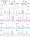
a) General structures with proton labels of free nanorings and their bimolecular aggregates. b–e) 1H and 1H DOSY spectra (500 MHz, CDCl3, 25 °C) of c-P12t-Bu, c-P8t-Bu, c-P12OOct, and c-P8OOct in the presence and absence of pyridine. * = residual solvent signal, Py = pyridine.
Figure 5
Figure 5
Comparative plots of HSQC cross-peaks for c-P12t-Bu, c-P8t-Bu, c-P12OOct, and c-P8OOct in the presence (pink) and absence of pyridine (green). The grouping of cross-peaks measured for aggregates (green) around cross-peaks measured for their free rings in the presence of pyridine (pink) was used to correlate between different types of protons in the free rings and their aggregates. 1H signals of “a” type β-pyrrole protons in all aggregates are broadened out beyond the limit of detection; thus, only the point for the free rings in the presence of pyridine is shown. Similarly, “b” type resonances are not observable in (c-P8t-Bu)2, only in the corresponding free ring.
Figure 6
Figure 6
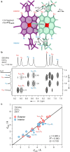
1H–1H NOESY analysis of (c-P12t-Bu)2. a) A 1/12th fragment of the model with the key calculated intermolecular distances used to distinguish between the exterior (e)/interior (i) and up (u)/down (d) proton environments. Hydrogen atoms of the t-Bu groups are omitted for clarity. b) Region of the NOESY spectrum highlighting key differences in NOE magnitudes for the assignment. c) Calculated versus experimental distances for the (c-P12t-Bu)2 aggregate.
Figure 7
Figure 7
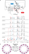
Variable-temperature 1H NMR spectra of the bimolecular aggregate of c-P12t-Bu. a) Structure of (c-P12t-Bu)2 with proton labeling at −50 °C and 1H NMR spectra from +50 °C to −50 °C, recorded at 500 MHz in CDCl3. * = residual solvent signal; x = CDCl3 satellite signals; Ex = exchange, detected by 1H–1H EXSY (ROESY) spectroscopy. b) Cartoon representation of the ring-on-ring rotation process in (c-P12t-Bu)2 leading to fast exchange of β-pyrrole protons at +50 °C, and slow exchange at −50 °C.
Figure 8
Figure 8
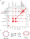
a) 1H–1H EXSY (NOESY) spectrum of a 2.2:1.0 mixture of (c-P8OOct)2 and c-P8OOct·Py8 showing direct exchange correlations between the two states (500 MHz, CDCl3, 25 °C, tmix = 200 ms). b) Cartoon representation of the equilibrium between c-P8OOct·Py8 and (c-P8OOct)2 giving rise to exchange correlations in 2D EXSY. Key exchange correlations between disaggregated ring and aggregate include: aai, aae, ooiu, ooeu, ooid, ooed, ppi, and ppe.
Figure 9
Figure 9
a) Structure of (c-P8OOct)2 with arrows indicating three internal rotation processes: interior aryl rotation (blue arrow), porphyrin rotation (green arrow), and exterior aryl rotation (red arrow), identified based on characteristic proton exchanges listed below. b) From top to bottom: selective 1H EXSY NMR spectra (CDCl3, 500 MHz, 25 °C) at different mixing times for targeted protons pi (tmix 2.4–200 ms), oiu (tmix 2.4–220 ms), ciu (tmix 2.4–220 ms), and 1H NMR spectrum of (c-P8OOct)2.
Figure 10
Figure 10
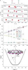
Magnetic field dependence of the aryl side chain 1H–13C total splittings (1TCH) for the (c-P12t-Bu)2 bimolecular aggregate. a) 13C-coupled HSQC spectra for ortho-aryl resonances oed and oid, showing a field-dependence of 1TCH resulting from a dipolar coupling contribution. b) Dependence of the observed 1TCH on the square of the magnetic field strength, plotted for both ortho- and para-aryl side chain protons. c) Root-mean-square deviation between experimental and calculated RDCs for model geometries of (c-P12t-Bu)2 with varying degrees of porphyrin rotation, given as the angle θ between the plane of the ring (mean plane of 12 Zn atoms) and the plane of the porphyrin units (24-atom mean plane).
Figure 11
Figure 11
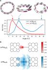
Molecular dynamics simulations of (c-P12t-Bu)2 and (c-P8OOct)2. a) Distribution of angles (as defined in Figure 10c) between porphyrin units and the plane of the nanoring defined by all zinc atoms, together with representative geometries for some of the most populated angles. Side chains and hydrogen atoms are omitted for clarity. b) Distribution of porphyrin zinc atom positions in one ring projected onto the plane of the other ring of the aggregate.

References

    1. Hartl F. U.; Hayer-Hartl M. Molecular chaperones in the cytosol: from nascent chain to folded protein. Science 2002, 295, 1852–1858. 10.1126/science.1068408. - DOI - PubMed
    1. Dobson C. M. Protein folding and misfolding. Nature 2003, 426, 884–890. 10.1038/nature02261. - DOI - PubMed
    1. Louros N.; Schymkowitz J.; Rousseau F. Mechanisms and pathology of protein misfolding and aggregation. Nat. Rev. Mol. Cell Biol. 2023, 24, 912–933. 10.1038/s41580-023-00647-2. - DOI - PubMed
    1. Spano F. C.; Silva C. H- and J-aggregate behavior in polymeric semiconductors. Annu. Rev. Phys. Chem. 2014, 65, 477–500. 10.1146/annurev-physchem-040513-103639. - DOI - PubMed
    1. Brédas J.-L.; Beljonne D.; Coropceanu V.; Cornil J. Charge-transfer and energy-transfer processes in π-conjugated oligomers and polymers: A molecular picture. Chem. Rev. 2004, 104, 4971–5004. 10.1021/cr040084k. - DOI - PubMed