. 2024 Nov:133:116-133.
doi: 10.1016/j.matbio.2024.08.007. Epub 2024 Aug 24.

Hyperglycemic environments directly compromise intestinal epithelial barrier function in an organoid model and hyaluronan (∼35 kDa) protects via a layilin dependent mechanism

Affiliations

Hyperglycemic environments directly compromise intestinal epithelial barrier function in an organoid model and hyaluronan (∼35 kDa) protects via a layilin dependent mechanism

Samreen Jatana et al. Matrix Biol. 2024 Nov.

Abstract

Background: Metabolic syndrome and diabetes in obese individuals are strong risk factors for development of inflammatory bowel disease (IBD) and colorectal cancer. The pathogenic mechanisms of low-grade metabolic inflammation, including chronic hyperglycemic stress, in disrupting gut homeostasis are poorly understood. In this study, we sought to understand the impact of a hyperglycemic environment on intestinal barrier integrity and the protective effects of small molecular weight (35 kDa) hyaluronan on epithelial barrier function.

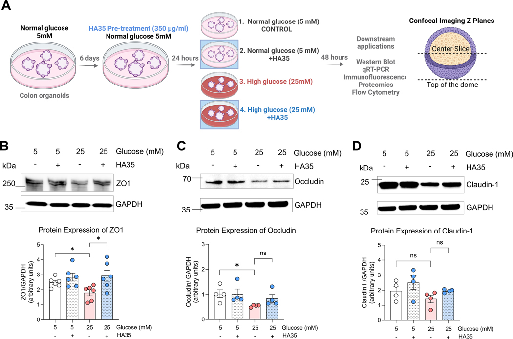
Methods: Intestinal organoids derived from mouse colon were grown in normal glucose media (5 mM) or high glucose media (25 mM) to study the impact of hyperglycemic stress on the intestinal barrier. Additionally, organoids were pretreated with 35 kDa hyaluronan (HA35) to investigate the effect of hyaluronan on epithelial barrier under high glucose stress. Immunoblotting as well as confocal imaging was used to understand changes in barrier proteins, quantitative as well as spatial distribution, respectively. Alterations in barrier function were measured using trans-epithelial electrical resistance and fluorescein isothiocyanate flux assays. Untargeted proteomics analysis was performed to elucidate mechanisms by which HA35 exerts a protective effect on the barrier. Intestinal organoids derived from receptor knockout mice specific to various HA receptors were utilized to understand the role of HA receptors in barrier protection under high glucose conditions.

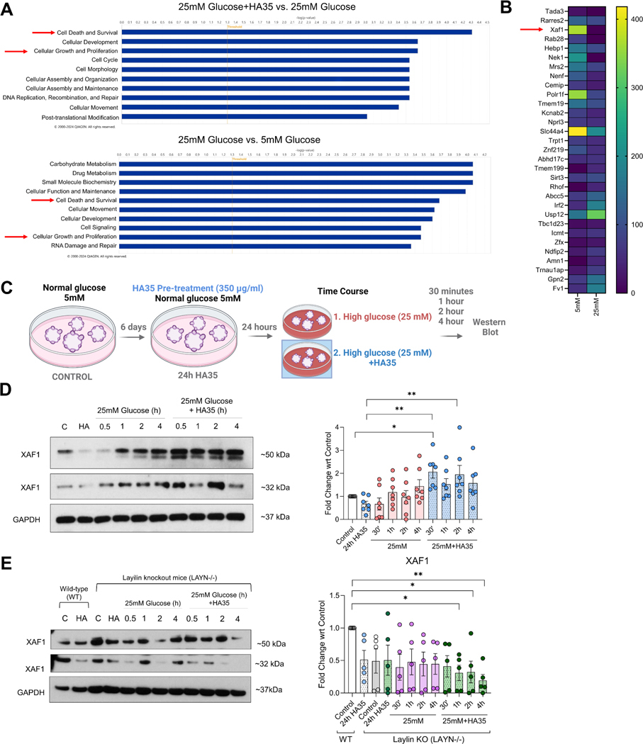
Results: We found that high glucose stress decreased the protein expression as well as spatial distribution of two key barrier proteins, zona occludens-1 (ZO-1) and occludin. HA35 prevented the degradation or loss of ZO-1 and maintained the spatial distribution of both ZO-1 and occludin under hyperglycemic stress. Functionally, we also observed a protective effect of HA35 on the epithelial barrier under high glucose conditions. We found that HA receptor, layilin, was involved in preventing barrier protein loss (ZO-1) as well as maintaining spatial distribution of ZO-1 and occludin. Additionally, proteomics analysis showed that cell death and survival was the primary pathway upregulated in organoids treated with HA35 under high glucose stress. We found that XIAP associated factor 1 (Xaf1) was modulated by HA35 thereby regulating apoptotic cell death in the intestinal organoid system. Finally, we observed that spatial organization of both focal adhesion kinase (FAK) as well as F-actin was mediated by HA35 via layilin.

Conclusion: Our results highlight the impact of hyperglycemic stress on the intestinal barrier function. This is of clinical relevance, as impaired barrier function has been observed in individuals with metabolic syndrome. Additionally, we demonstrate barrier protective effects of HA35 through its receptor layilin and modulation of cellular apoptosis under high glucose stress.

Keywords: Apoptosis; Hyaluronan; Hyperglycemia; Intestinal barrier; Layilin; Metabolic syndrome; Organoids.


Conflict of interest statement

Conflict of interest The authors declare that they have no known competing financial interests or personal relationships that could have appeared to influence the work reported in this paper.

Figures

Fig. 1.
Fig. 1.
HA35 protects against the effects of hyperglycemia on tight junction protein expression. (A) Schematic showing in vitro set up for organoid assays. Murine distal colon organoids were grown and expanded in media containing normal glucose (5 mM) for 6 days. Prior to addition of high glucose (25 mM), organoids were pre-treated with HA35 (350 μg/ml) for 24 h. Organoids were collected for downstream assays 48 h after addition of high glucose (25 mM) except for time course studies. Confocal images were taken either at the center of the dome (center slice) or the top of the organoid domes (B) Western blots and bar plots showing the quantification of ZO-1, occludin, and claudin-1 proteins in colonic organoids across four treatment groups (48 h post-treatment). N = 4–6 wild-type (WT) mouse organoids. Results are shown as Mean ± SEM. *p < 0.05, one-way ANOVA in comparison to 5 mM control group and 25 mM high glucose group (Dunnett’s multiple comparison test).
Fig. 2.
Fig. 2.
HA35 pre-treatment maintains spatial distribution of ZO-1 and occludin under high glucose conditions. (A) Representative immunofluorescence (IF) images showing ZO-1 (red) and E-cadherin (green) staining in mouse colon organoids. Bottom panel: magnification of white boxes in top panel (inset). Images acquired at the center slice of spheroid dome. (B) Representative IF images showing occludin (red) and E-cadherin (green) staining in mouse colon organoids. Bottom panel: magnification of white boxes in top panel (inset). Images acquired at the top of the spheroid dome. (C) Representative IF images showing claudin-1 (magenta) and E-cadherin (yellow) in mouse colon organoids. Bottom panel: magnification of white boxes in top panel (inset). Images acquired at the center slice of spheroid dome. DAPI marks nuclei in blue. All images were acquired using confocal microscopy. Top panels were acquired using 40X oil objective and insets (bottom panels) using 63X oil objective. Representative images from N = 3 WT mouse organoids, 48 h post-treatment. Scale bar=20 μm (Inset=5 μm).
Fig. 3.
Fig. 3.
HA35 protects functional barrier integrity under hyperglycemic stress. (A) Representative images from mouse colon “flipped apical surface out” organoids pre-treated with HA35 followed by addition of high glucose (25 mM). 40 kDa FITC-dextran was added to visualize outside-in (apical to basolateral) flux to measure barrier function. Organoids with dark centers indicative of reduced FITC-dextran transit. Alternatively, organoid centers that were fluorescent green were indicative of impaired barrier or increased FITC transit (black arrows). Images acquired using confocal microscopy (40X oil objective). Scale bar=20 μm. (B) Bar plot showing the number of organoids per field containing dark centers. N = 3 WT mouse organoids, n = 3 images acquired per treatment group, 48 h post-treatment with high glucose (25 mM) and 10 min post-treatment with 40 kDa FITC-dextran. *p < 0.05, ****p < 0.0001, one-way ANOVA (inter group analysis, post-hoc Tukey). (C) Transepithelial electrical resistance (TEER) measured in monolayers across the four treatment groups over a period of 4 days. TEER values were normalized to Day 0 measurements (start of the experiment). *p < 0.05, unpaired t-Test with Welch’s correction. Monolayers were grown from N = 5 WT mouse organoids.
Fig. 4.
Fig. 4.
HA receptor, layilin, is involved in the maintenance of barrier integrity under hyperglycemic stress. (A) Representative western blot and quantification (densitometry) of protein expression of ZO-1 in LAYN−/− organoid lysates across the four treatment groups. N = 6 LAYN−/− mouse organoids, 48 h post-treatment, ***p < 0.001, one-way ANOVA (in comparison to 5 mM control and 25 mM high glucose, Dunnett’s multiple comparison test). (B) Representative western blot and quantification (densitometry) of protein expression of occludin in LAYN−/− organoid lysates across the four treatment groups. N = 6 LAYN−/− mouse organoids, 48 h post-treatment. (C) Representative IF images showing ZO-1 (red) staining in LAYN−/− organoids across the four treatment groups. Top panel shows images acquired at the top of organoid domes and bottom panels show images acquired at the center slice. (D) Representative IF images showing occludin (red) and E-cadherin (green) staining in LAYN−/− mouse organoids across the four treatment groups. Images were obtained at the top of the organoid domes. All IF images were acquired using a confocal microscope (40X oil objective), representative N = 3 LAYN−/− mouse organoids, 48 h post-treatment. Scale bar=20 μm.
Fig. 5.
Fig. 5.
HA35 regulates cell death and survival under high glucose stress. (A) Pathway analysis using IPA software to identify top cellular pathways upregulated in 25 mM glucose+HA35 vs. 25 mM glucose (top) and 25 mM glucose vs. 5 mM glucose treatment groups (bottom). Threshold set at 1.3 -log (p-value). N = 4 WT mouse organoids, 48 h post-treatment. (B) Heatmaps generated from proteomics data showing changes (up- and downregulated) in proteins that were significantly up- or downregulated 48 h post-treatment in high glucose (25 mM) treatment vs. normal glucose controls (5 mM), p < 0.05. (C) Schematic showing in vitro organoid set up for western blot time course experiments. Samples were collected 30 min, 1 h, 2 h and 4 h post-treatment. (D) Representative western blot and quantification (densitometry) of protein expression of Xafl in organoid lysates across the four treatment groups. Xaf1 has two separate bands detected at ~50 kDa and ~32 kDa size. N = 6 WT mouse organoids utilized for western blotting. *p < 0.05, **p < 0.01, one-way ANOVA (analyses performed in comparison to 5 mM control as well as 24 h HA35 treatment control, Dunnett’s multiple comparison test). (E) Representative western blots and quantification (densitometry) of protein expression of Xafl in LAYN−/− organoid lysates across four treatment groups. First two lanes in the gel are WT organoids grown in 5 mM media control (C) and WT organoids treated with HA35 for 24 h. N = 5 WT and LAYN−/− mouse organoids utilized for western blotting, *p < 0.05 in comparison to WT control (C), one-way ANOVA (Dunnett’s multiple comparison test).
Fig. 6.
Fig. 6.
HA35 counteracts the effect of hyperglycemic stress apoptotic cell death. (A) Representative IF images showing cleaved caspase-3 (red) and E-cadherin (green) staining in WT organoids. DAPI are marked in blue. Representative images from N = 3 WT mouse organoids. Scale bar=20 μm. (B) Bar plot showing percentage of apoptotic cells measured by flow cytometry. N = 5 WT mouse organoids. *p < 0.05, one-way ANOVA, inter-group analysis performed in comparison to 5 mM control and 25 mM high glucose treatment (Dunnett’s multiple comparison test). (C) Representative IF images showing cleaved caspase-3 (red) and E-cadherin (green) staining in LAYN−/− organoids. DAPI are marked in blue. Representative images from N = 3 LAYN−/− mouse organoids. Scale bar=20 μm. (D) Bar plot showing percentage of apoptotic cells measured by flow cytometry. N = 4 LAYN−/− mouse organoids. *p < 0.05, one-way ANOVA, inter-group analysis performed in comparison to control (Dunnett’s multiple comparison test).
Fig. 7.
Fig. 7.
HA35 maintains spatial distribution of F-actin and focal adhesion kinase (FAK) under high glucose conditions. (A) Representative IF images showing ZO-1 (red), F-actin (phalloidin, magenta), and focal adhesion kinase (FAK, green) staining in WT organoids. DAPI are marked in blue. Representative images from N = 3 WT mouse organoids. Scale bar=20 μm. (B) FAK staining (green) inset from regions highlighted in white box (panel A). Scale bar=5 μm. All images are acquired at the center slice of the organoid dome.
Fig. 8.
Fig. 8.
HA35 maintains spatial distribution of F-actin and focal adhesion kinase (FAK) under high glucose conditions via layilin. (A) Representative IF images showing ZO-1 (red), F-actin (phalloidin, magenta), and focal adhesion kinase (FAK, green) staining in LAYN−/− organoids. DAPI are marked in blue. Representative images from N = 3 LAYN−/− mouse organoids. Scale bar=20 μm. (B) FAK staining (green) inset from regions highlighted in white box (panel A). Scale bar=5 μm. All images are acquired at the center slice of the organoid dome.
Fig. 9.
Fig. 9.
Schematic summarizing findings from the study. In this study, we showed that hyperglycemic stress increases protein loss (ZO-1) and spatial distribution (ZO-1, occludin) of key intestinal barrier proteins in a mouse intestinal organoid model. Small molecular weight hyaluronan (35 kDa), HA35, plays a protective effect on barrier function under high glucose conditions via layilin. HA35 regulates apoptotic cell death in organoids grown under hyperglycemic stress by modulating the levels of Xaf1 protein. HA35 maintains the spatial arrangement of FAK and F-actin under hyperglycemic stress. The effects of HA35 on the intestinal barrier function are likely regulated by its receptor layilin via the layilin-integrin-FAK axis.

References

    1. Schleh MW, Caslin HL, Garcia JN, Mashayekhi M, Srivastava G, Bradley AB, Hasty AH, Metaflammation in obesity and its therapeutic targeting, Sci. Transl. Med 15 (723) (2023) eadf9382. - PMC - PubMed
    1. Hotamisligil GS, Inflammation, metaflammation and immunometabolic disorders, Nature 542 (7640) (2017) 177–185. - PubMed
    1. Collaboration NCDRF, Worldwide trends in underweight and obesity from 1990 to 2022: a pooled analysis of 3663 population-representative studies with 222 million children, adolescents, and adults, Lancet 403 (10431) (2024) 1027–1050. - PMC - PubMed
    1. Priest C, Tontonoz P, Inter-organ cross-talk in metabolic syndrome, Nat. Metab 1 (12) (2019) 1177–1188. - PubMed
    1. Ma B, Wang X, Ren H, Li Y, Zhang H, Yang M, Li J, High glucose promotes the progression of colorectal cancer by activating the BMP4 signaling and inhibited by glucagon-like peptide-1 receptor agonist, BMC Cancer 23 (1) (2023) 594. - PMC - PubMed

LinkOut - more resources