

Inflammatory signature in amyotrophic lateral sclerosis predicting disease progression
- PMID: 39187524
- PMCID: PMC11347586
- DOI: 10.1038/s41598-024-67165-9
Inflammatory signature in amyotrophic lateral sclerosis predicting disease progression
Abstract
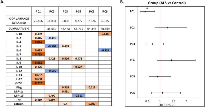
Experimental studies identified a role of neuroinflammation in the pathogenesis of neurodegenerative diseases, including amyotrophic lateral sclerosis (ALS). However, the role of inflammatory molecules as diagnostic and prognostic biomarkers in patients with ALS is unclear. In this cross-sectional study, the cerebrospinal fluid (CSF) levels of a set of inflammatory cytokines and chemokines were analyzed in 56 newly diagnosed ALS patients and in 47 age- and sex-matched control patients without inflammatory or degenerative neurological disorders. The molecules analyzed included: interleukin (IL)-1β, IL-2, IL-4, IL-5, IL-6, IL-7, IL-8, IL-9, IL-10, IL-12, IL-13, IL-17, granulocyte colony stimulating factor (GCSF), macrophage inflammatory protein (MIP)-1a, MIP-1b, tumor necrosis factors (TNF), eotaxin. Principal component analysis (PCA) was used to explore possible associations between CSF molecules and ALS diagnosis. In addition, we analyzed the association between CSF cytokine profiles and clinical characteristics, including the disease progression rate score, and peripheral inflammation assessed using the Neutrophil-to-lymphocyte ratio (NLR). PCA identified six principal components (PCs) explaining 70.67% of the total variance in the CSF cytokine set. The principal component (PC1) explained 26.8% of variance and showed a positive load with CSF levels of IL-9, IL-4, GCSF, IL-7, IL-17, IL-13, IL-6, IL-1β, TNF, and IL-2. Logistic regression showed a significant association between PC1 and ALS diagnosis. In addition, in ALS patients, the same component was significantly associated with higher disease progression rate score and positively correlated with NLR. CSF inflammatory activation in present in ALS at the time of diagnosis and may characterize patients at higher risk for disease progression.
Keywords: Amyotrophic lateral sclerosis (ALS); Cerebrospinal fluid (CSF); Cytokines; Disease progression; Neuroinflammation; Neutrophil-to-lymphocytes ratio (NLR).
© 2024. The Author(s).
Conflict of interest statement
FB acted as advisory board members for Teva and Roche and received honoraria for speaking or consultation fees from Merck Serono, Teva, Biogen Idec, Sanofi, and Novartis, and non-financial support from Merck Serono, Teva, Biogen Idec, and Sanofi. DC is an Advisory Board member of Almirall, Bayer Schering, Biogen, GW Pharmaceuticals, Merck Serono, Novartis, Roche, Sanofi-Genzyme, and Teva and received honoraria for speaking or consultation fees from Almirall, Bayer Schering, Biogen, GW Pharmaceuticals, Merck Serono, Novartis, Roche, Sanofi-Genzyme, and Teva. He is also the principal investigator in clinical trials for Bayer Schering, Biogen, Merck Serono, Mitsubishi, Novartis, Roche, Sanofi-Genzyme, and Teva. His preclinical and clinical research was supported by grants from Bayer Schering, Biogen Idec, Celgene, Merck Serono, Novartis, Roche, Sanofi- Genzyme and Teva. The other authors declare that the research was conducted in the absence of any commercial or financial relationships that could be construed as a potential conflict of interest.
Figures


References
MeSH terms
Substances
LinkOut - more resources
Full Text Sources
Medical
Research Materials
Miscellaneous
