. 2024 Aug 26;30(1):132.
doi: 10.1186/s10020-024-00897-6.

Secretome of brain microvascular endothelial cells promotes endothelial barrier tightness and protects against hypoxia-induced vascular leakage

Affiliations

Secretome of brain microvascular endothelial cells promotes endothelial barrier tightness and protects against hypoxia-induced vascular leakage

Rodrigo Azevedo Loiola et al. Mol Med. .

Abstract

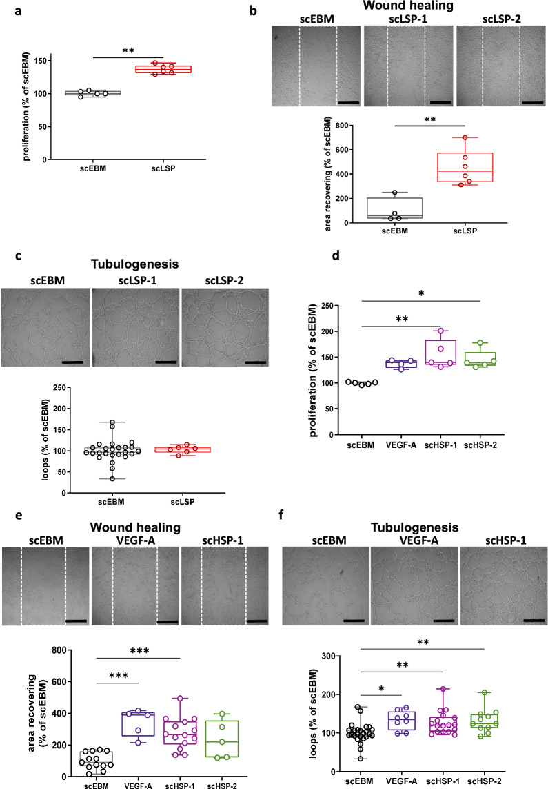
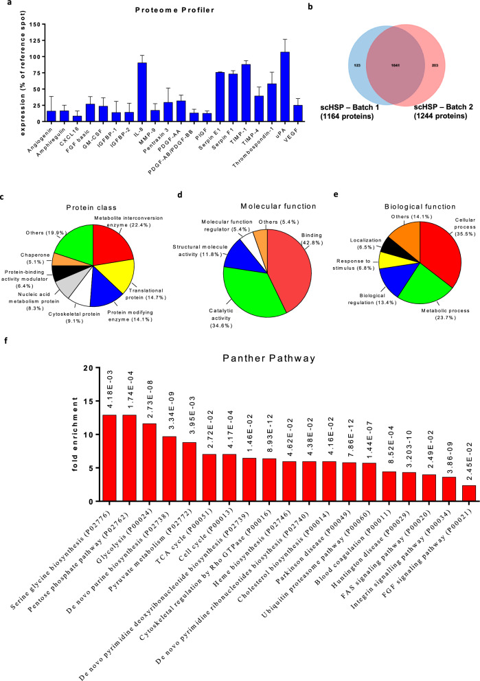
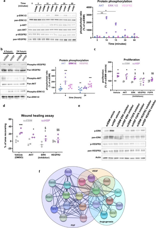
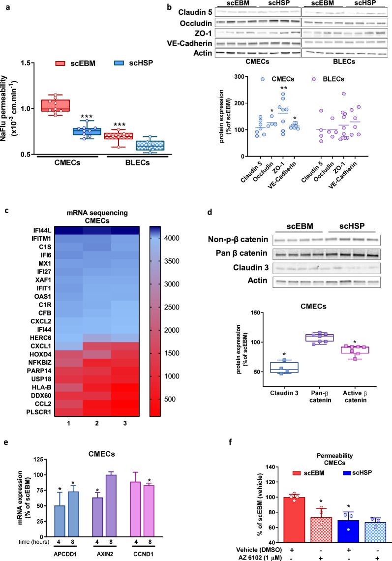
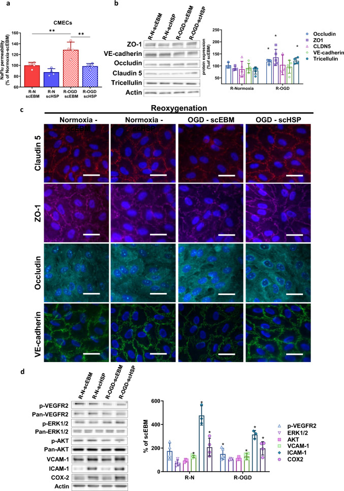
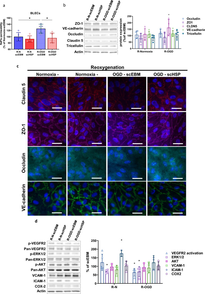
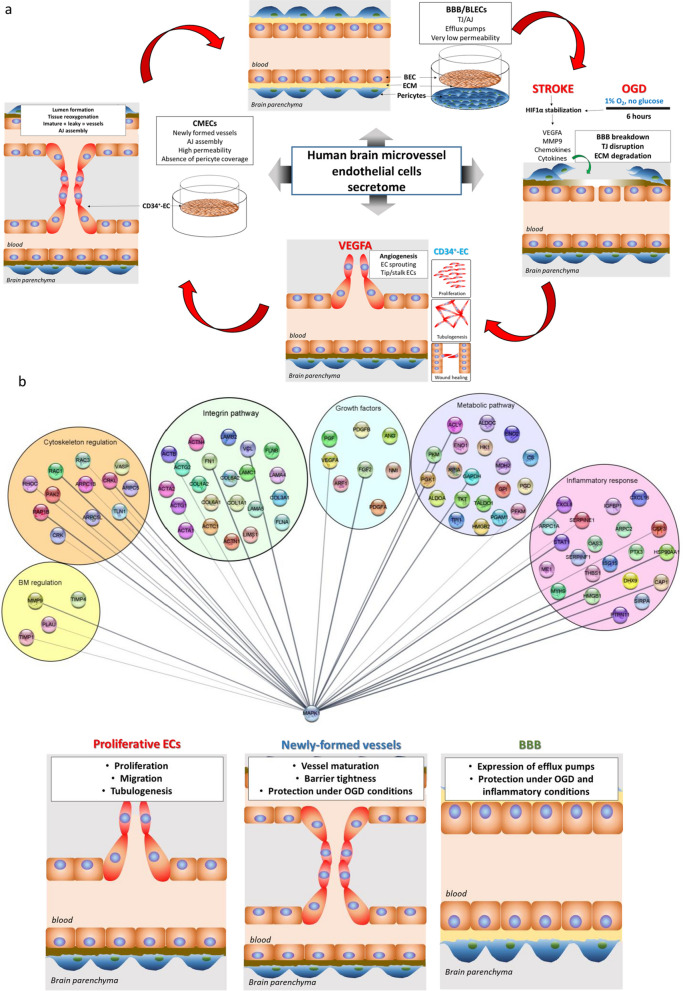
Cell-based therapeutic strategies have been proposed as an alternative for brain and blood vessels repair after stroke, but their clinical application is hampered by potential adverse effects. We therefore tested the hypothesis that secretome of these cells might be used instead to still focus on cell-based therapeutic strategies. We therefore characterized the composition and the effect of the secretome of brain microvascular endothelial cells (BMECs) on primary in vitro human models of angiogenesis and vascular barrier. Two different secretome batches produced in high scale (scHSP) were analysed by mass spectrometry. Human primary CD34+-derived endothelial cells (CD34+-ECs) were used as well as in vitro models of EC monolayer (CMECs) and blood-brain barrier (BBB). Cells were also exposed to oxygen-glucose deprivation (OGD) conditions and treated with scHSP during reoxygenation. Protein yield and composition of scHSP batches showed good reproducibility. scHSP increased CD34+-EC proliferation, tubulogenesis, and migration. Proteomic analysis of scHSP revealed the presence of growth factors and proteins modulating cell metabolism and inflammatory pathways. scHSP improved the integrity of CMECs, and upregulated the expression of junctional proteins. Such effects were mediated through the activation of the interferon pathway and downregulation of Wnt signalling. Furthermore, OGD altered the permeability of both CMECs and BBB, while scHSP prevented the OGD-induced vascular leakage in both models. These effects were mediated through upregulation of junctional proteins and regulation of MAPK/VEGFR2. Finally, our results highlight the possibility of using secretome from BMECs as a therapeutic alternative to promote brain angiogenesis and to protect from ischemia-induced vascular leakage.

Keywords: Angiogenesis; Blood–brain barrier; Brain microvascular endothelial cells; Cardiovascular disease; Cell therapy; Regenerative medicine; Secretome; Stroke.


Conflict of interest statement

PB, AK and MM are employees of Pure Biologics S.A. Authors report no competing interests.

Figures

Fig. 1
Fig. 1
BMEC-secretome promotes in vitro angiogenesis. BMEC-secretomes under low scale production conditions (scLSP) promote CD34+-ECs proliferation (a), migration (b), but not tubulogenesis (c). Results were obtained with six different batches of scLSP (scLSP-1 to scLSP-6). Representative pictures are from scLSP-1 and scLSP-2. As indicated in the material and methods section, Wilcoxon–Mann–Whitney test has been used for ac. Further, BMEC-secretome produced at high-scale conditions (scHSP) increased CD34+-ECs proliferation (d), migration (e), and tubulogenesis (f). Besides, two scHSP batches were compared and showed similar effects (scHSP-1, batch 1; sHSP-2, batch 2). The effects of scHSP were similar to those observed with VEGF-A (50 ng/mL). Kruskal–Wallis test has been used for df. Scale bar: 200 μm
Fig. 2
Fig. 2
Identification of proteins in scHSP. Detection of pro-angiogenesis factors in two batches of scHSP was performed by Angiogenesis Proteome Profiler (a). Two different batches of scHSP were analysed by mass spectrometry (DDA-MS method). Over 1200 proteins were identified and the two batches of scHSP shared 1041 proteins (b). The set of proteins shared between two batches of scHSP (1041) were converted in gene identifiers and then submitted to an enrichment analysis using the GeneOntology/Panther software. The proteins were classified according to protein class (c), molecular (d), or biological function (e). Among the Panther pathways that were over-represented in the enrichment analysis of scHSP samples, several were related to cell metabolism (f)
Fig. 3
Fig. 3
scHSP promotes in vitro angiogenesis mediated by MAPK and VEGFR2 activation. scHSP induces a rapid and consistent increase in ERK1/2 phosphorylation, whereas it does not affect AKT or VEGFR2 activation (a). ERK1/2 activation remains until 4 h after treatment, while VEGFR2 phosphorylation is upregulated after 24 h of treatment (b). Pre-incubation with U0126 (MEK/ERK inhibitor—1 μmol/L), VEGFR2 kinase inhibitor VII (10 μmol/L) and FGFR tyrosine kinase inhibitor (1 μmol/L) reduced the scHSP-induced proliferation (c) while VEGFR2 and FGFR inhibition reduced the scHSP-induced migration (d). Western blot detection of ERK1/2 and VEGFR2 phosphorylation on cells treated with either vehicle (DMSO), MEK/ERK1/2, or VEGFR2, or FGFR inhibitors (e). Proteins classified as involved in angiogenesis (Hossein Geranmayeh et al. 2023), VEGF-A (Gosselet et al. 2021), and FGF (Maki et al. 2018) were grouped, then converted in gene identifiers and an interaction network was prepared using Cytoscape software (version 3.8.2, released 2020.10.24). Such pathways share in common the protein MAPK1 (ERK2) (f). Data represents median with individual data (n = 3–7), *p < 0.05, **p < 0.01, ***p < 0.001, vs scEBM for all graphs; and $p < 0.05, $$p < 0.01, vs scHSP vehicle for c and d
Fig. 4
Fig. 4
scHSP promotes endothelial barrier tightness. scHSP reduced the permeability of CMECs, while it did not affect BLECs (a). One way ANOVA was performed with post hoc Tukey’s multiple comparisons test, versus scEBM CMECs. scHSP up-regulated expression of junctional proteins on CMECs (b). Results represent Scatter dot plot. Individual data and median. Heat map representing 23 genes up-regulated by scHSP on CMECs, most of them associated with the IFN pathway (c). scHSP reduced the content of active β-catenin and claudin 3 (d) and downregulated mRNA expression of Wnt pathway targets APCDD1, Axin2, and CCND1 on CMECs (e). Co-incubation with Wnt inhibitor (AZ6102, 1 μmol/L) decreased barrier permeability of cells treated with scEBM (f)
Fig. 5
Fig. 5
OGD-induced vascular leakage in vitro is prevented by scHSP. Treatment with scHSP during reoxygenation (24 h) abolished the OGD-induced increase of permeability on CMECs (a). Results represent separated bar graph with means ± SD (One way ANOVA followed by Post hoc Tukey’s multiple comparisons test versus R-OGD scEBM condition). WB analysis revealed that scHSP increased occludin, ZO-1, and tricellulin expression (b) and favored their localization at cell junctions (c). scHSP increased VEGFR2 activation, while it did not affect ERK1/2 or AKT activation (d). Besides, scHSP up-regulated VCAM-1, ICAM-1, and COX2 expression (d). As indicated in the material and method section, data represent median (interquartile range) (for b) or mean ± SD (for c). Scale bar: 10 μm
Fig. 6
Fig. 6
OGD-induced in vitro BBB leakage is prevented by scHSP. Treatment with scHSP during reoxygenation (24 h) abolished the OGD-induced increase of permeability on BLECs (a). Results show means ± SD, One way ANOVA and Post hoc Holm-Šídák’s multiple comparisons test, versus R-OGD scEBM condition. WB analysis revealed that scHSP increased claudin 5 expression (b) and favored its localization at the cell junctions (c). scHSP decreased VEGFR2 and ERK1/2 activation during reoxygenation (d). Moreover, scHSP increased ICAM-1 expression in normoxic conditions but had no effect on BLECs exposed to OGD (d). Data represent means ± SD, versus scEBM conditions. Scale bar: 10 μm
Fig. 7
Fig. 7
Network map of potential proteins/pathways involved in scHSP-induced effects. a Schematic overview of angiogenesis process and in vitro assays proposed for evaluating EPC-secretome effects. Human CD34+ derived cord-blood hematopoietic endothelial cells (CD34+-ECs) were used to study angiogenesis (proliferation, cell migration, and tubulogenesis). To investigate mechanisms involved in vascular maturation, CD34+-EC were seeded on the surface of Matrigel™-coated Transwell inserts and cultivated in monocultures (CMECs). Finally, to study the effect of EPC-secretome on brain capillaries, we used an in vitro BBB model which consisted in seeding CD34+-ECs on Matrigel™-coated Transwell inserts and co-cultivating them with human brain pericytes, which enabled CD34+-ECs to acquire a brain-like endothelial cell phenotype (BLECs). b Proteins present in scHSP were converted in gene identifiers and were then grouped according to their biological function/pathway (Metabolic pathways, Growth factors, Inflammatory response, basal membrane (BM) regulation, cytoskeletal regulation, and integrin pathway). An interaction network was prepared using the Cytoscape software (version 3.8.2, released 2020.10.24). The regulation of MAPK activity might play an essential role in the scHSP-induced angiogenesis in human primary ECs. In parallel, scHSP promotes vascular maturation of newly-formed vessels and protects the BBB integrity under ischemic and inflammatory conditions

References

    1. Abbott NJ, Ronnback L, Hansson E. Astrocyte-endothelial interactions at the blood–brain barrier. Nat Rev Neurosci. 2006;7(1):41–53. 10.1038/nrn1824 - DOI - PubMed
    1. Abdullahi W, Tripathi D, Ronaldson PT. Blood–brain barrier dysfunction in ischemic stroke: targeting tight junctions and transporters for vascular protection. Am J Physiol Cell Physiol. 2018;315(3):C343–56. 10.1152/ajpcell.00095.2018 - DOI - PMC - PubMed
    1. Akarasereenont P, Techatraisak K, Thaworn A, Chotewuttakorn S. The expression of COX-2 in VEGF-treated endothelial cells is mediated through protein tyrosine kinase. Mediat Inflamm. 2002;11(1):17–22. 10.1080/09629350210311 - DOI - PMC - PubMed
    1. Arai K, Jin G, Navaratna D, Lo EH. Brain angiogenesis in developmental and pathological processes: neurovascular injury and angiogenic recovery after stroke. FEBS J. 2009;276(17):4644–52. 10.1111/j.1742-4658.2009.07176.x - DOI - PMC - PubMed
    1. Bai Y-Y, Wang L, Chang D, Zhao Z, Lu C-Q, Wang G, et al. Synergistic effects of transplanted endothelial progenitor cells and RWJ 67657 in diabetic ischemic stroke models. Stroke. 2015;46(7):1938–46. 10.1161/STROKEAHA.114.008495 - DOI - PubMed

LinkOut - more resources