Case Reports
. 2024 Aug 27;25(1):805.
doi: 10.1186/s12864-024-10478-w.

A novel deletion in the BLOC1S6 Gene Associated with Hermansky-Pudlak syndrome type 9 (HPS-9)

Affiliations
Case Reports

A novel deletion in the BLOC1S6 Gene Associated with Hermansky-Pudlak syndrome type 9 (HPS-9)

Seyyed Mohammad Kahani et al. BMC Genomics. .

Abstract

Background: Hermansky-Pudlak Syndrome (HPS), a rare autosomal recessive disorder, is characterized by oculocutaneous albinism, bleeding diathesis, and sometimes severe lung problems and inflammatory bowel disease. Symptoms include skin and hair pigmentation variations, along with visual impairments. Variants in eleven genes encoding protein complexes essential for membrane trafficking and intracellular endosomal transport pathways underlie various recognized HPS subtypes. This study focuses on HPS-9, a subtype of Hermansky-Pudlak Syndrome caused by a variant in the BLOC1S6 gene, which is a subunit of the BLOC1 complex. In this study, a novel Copy Number Variation (CNV) in the aforementioned gene in an Iranian family is reported. The study aims to better understand the etiology of HPS-9 symptoms by identifying and confirming the variant and determining whether the gene is expressed despite the deletion. There have only been five reports of this syndrome in the literature thus far. Our novel CNV represents a significant contribution to understanding the genetic basis of HPS-9.

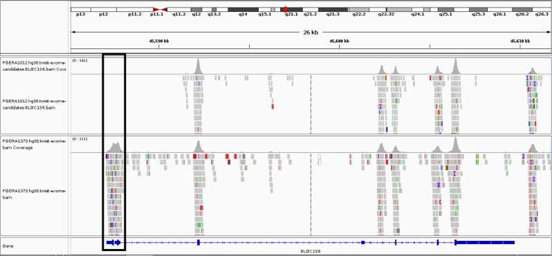
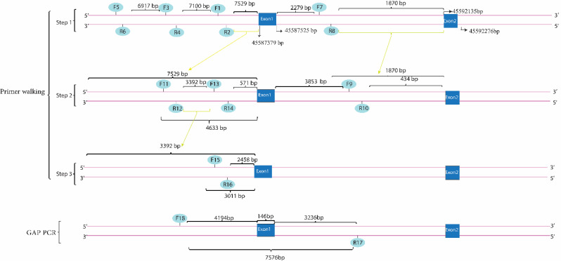
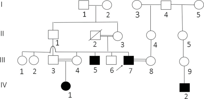
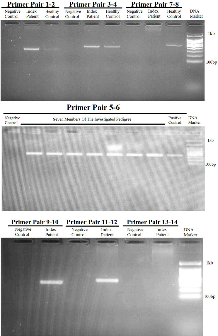
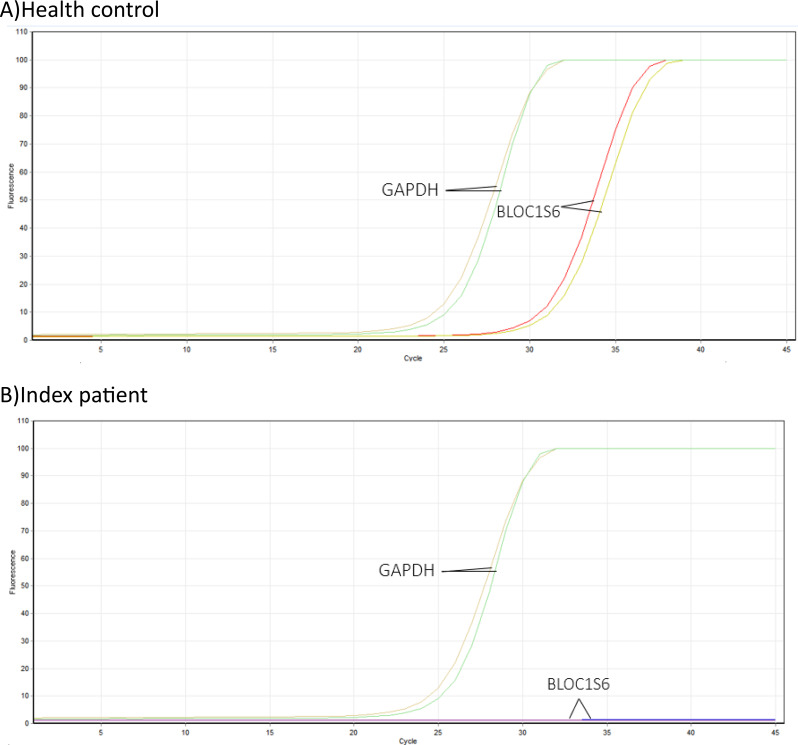
Results: This study investigates a male patient presenting with albinism. Whole Exome Sequencing (WES) identified a homozygous deletion of approximately 350 bp using CNV analysis. The deletion affects the intronic region of the BLOC1S6 gene, causing uncertainties in defining the exact boundaries due to WES limitations. Primer walking and GAP-PCR techniques were used to define the deletion boundaries. Subsequent assessments of this variant across other family members helped identify homozygous affected members and heterozygous carriers. The absence of BLOC1S6 expression in the affected individual was confirmed through Real-time PCR experiments. These findings underscore the importance of understanding the implications for the patient's healthcare and potential therapeutic approaches.

Conclusion: This study introduces a case of Hermansky-Pudlak Syndrome Type 9 (HPS-9) caused by a homozygous deletion in the BLOC1S6 gene. We identified an approximately 7-kb deletion encompassing exon 1 and the intronic region of the gene. The absence of BLOC1S6 expression, confirmed via Real-time PCR, highlights the importance of studying the pathogenicity of the deletion and its impact on the patient's health. Our findings contribute to the sparse knowledge on HPS-9 and underscore the need for further exploration into the genetic causes of this rare disorder.

Keywords: Albinism; GAP-PCR; Hermansky-pudlock syndrome type 9; Primer-walking; Whole exome sequencing.


Conflict of interest statement

The authors declare no competing interests.

Figures

Fig. 1
Fig. 1
Comparison of the BAM file of the proband with the BAM file of a healthy control individual. The upper row corresponds to the proband and the lower row corresponds to the control. The deletion is evident for the proband within the region specified. The figure was generated from the IGV software
Fig. 2
Fig. 2
Illustrates the primer walking process followed by GAP-PCR to identify the boundaries of a genomic deletion. The primer pairs (1–2, 3–4, 5–6) were designed to target regions upstream of the first exon of the BLOC1S6 gene, successfully amplifying their target regions. The breakpoint was determined using primer pair 11–12, which amplified the expected PCR product. Primer pairs 13–14 and 15–16 were designed to further delineate the deletion boundaries and were found to be within the deleted region. Primer pairs 7 and 8 were then designed for the downstream deletion boundary, and primer pair 9–10 was used to approach exon 2. The data from primer walking led to the hypothesis that primers 10 and 11 could be used for the GAP-PCR method. The resulting PCR product was subjected to Sanger sequencing, and based on the sequence, another reverse primer [15] was designed. The GAP-PCR method was applied using primers 11 and 17, which produced an 800-base-pair PCR product confirmed by gel electrophoresis. We performed bidirectional Sanger sequencing on the PCR product until the results were clear and free of noise. To achieve a 450-base-pair PCR product, We designed a forward primer 18, which was placed 439 bases downstream of primer 11. We then used this forward primer 18, along with reverse primer 17, through the GAP-PCR method. The corresponding reference genomic sequence for this region was found to be approximately 7 kb in length, and the GAP-PCR based test confirmed the presence of the deletion  (Please note that while the numerical scales indicated in this image, such as base pair (bp) measurements, are accurate and reflect true sizes of DNA fragments and genomic features, the physical dimensions of the image itself are not to scale)
Fig. 3
Fig. 3
The index case, highlighted by an arrow, was a 32-year-old male with parents who were distant relatives by consanguinity. His 68-year-old father, who remained healthy until his natural death, and his 58-year-old mother are notable family members. The proband married a 34-year-old woman, who also had a family history of distant consanguinity. Originating from a small village in Iran, this case is further intriguing due to the presence of the albinism phenotype not only in his brother and niece but also in the grandson of his grandfather-in-law. In addition to the index patient and his immediate family, it is noted that another distant relative appears to be unrelated to the proband. However, the presence of consanguinity issues within the family suggests that there may be other carriers and affected individuals residing in the village. This observation underscores the potential for a broader impact of genetic disorders within the community, highlighting the importance of further investigation to identify and understand the extent of the genetic predisposition among the population
Fig. 4
Fig. 4
Results of the gel electrophoresis after performing PCR using primer pairs targeting the flanking regions of exon 1 of the BLOC1S6 gene. It is evident, the results of PCR using all primer pairs targeting the upstream region of exon 1 of the BLOC1S6 gene, including primer pairs 1–2, 3–4, 5–6, and 11–12 are positive for the index patient (proband) and the healthy control. this indicates that the targeted loci are not deleted. However, the results for primer pair 13–14, that this pair targets a locus within the deleted region. On the other hand, the result of PCR using primer pair 9–10 targeting the downstream region of exon 1 of the BLOC1S6 is positive for the index patient and the healthy control, whereas the result of PCR using primer pair 7–8 used in the first try to target the downstream side was negative for the index patient, which indicates that this locus is located in the deletion region
Fig. 5
Fig. 5
Results of the gel electrophoresis after performing PCR using forward primer from upstream side and reverse primer from downstream side of exon 1 of the BLOC1S6 gene. To set up the GAP-PCR, we first approached the deletion region as closely as possible using primers 10 and 11. Subsequently, we moved closer to the deletion site, using primers 11 and 17. Finally, we obtained the smallest replicable fragment using primers 17 and 18
Fig. 6
Fig. 6
The melt curve analysis for the BLOC1S6 gene showed no peak, indicated by a horizontal dash at the bottom of the plot. This suggests that there is no alternative transcript of the BLOC1S6 gene that can be expressed, and therefore the BLOC1S6 gene is not expressed
Fig. 7
Fig. 7
The figure show the examination of the amplification curve for the BLOC1S6 gene revealed no peak, which is symbolized by a horizontal line at the base of the graph. This absence of a peak suggests that the BLOC1S6 gene does not have any alternative transcripts that can be expressed. Conversely, the amplification curve analysis for the GAPDH gene did show a peak. This is not surprising, as the GAPDH gene is a housekeeping gene that is typically found in blood cells. These findings suggest that the BLOC1S6 gene is not being expressed in the patient’s cells, while the GAPDH gene, a housekeeping gene, is being expressed. This could potentially impact the patient’s health condition, given that the patient has a deletion in exon 1 of the BLOC1S6 gene. The hypothesis was to investigate if there is an alternative transcript that can be expressed despite this deletion. The results indicate that there is no such alternative transcript, which could have significant implications for the patient’s health and treatment
Fig. 8
Fig. 8
Gel electrophoresis analysis revealing the absence of a product in patient samples compared to healthy control
Fig. 9
Fig. 9
The figure illustrates the positions of identified genetic variants within the exons of the BLOC1S6 gene. Male cases are represented by red circles, females by blue circles, and the star indicates the date of the study reporting these cases

References

    1. Grønskov K, Ek J, Brondum-Nielsen K. Oculocutaneous albinism. Orphanet J Rare Dis. 2007;2(1):43. 10.1186/1750-1172-2-43 - DOI - PMC - PubMed
    1. Moreno-Artero E, Morice-Picard F, Bremond-Gignac D, Drumare-Bouvet I, Duncombe-Poulet C, Leclerc-Mercier S, et al. Management of albinism: French guidelines for diagnosis and care. J Eur Acad Dermatol Venereol. 2021;35(7):1449–59. 10.1111/jdv.17275 - DOI - PubMed
    1. Dumitrescu AV, Pfeifer WL, Drack AV. Clinical phenocopies of albinism. J Aapos. 2021;25(4):220. e1-.e8.10.1016/j.jaapos.2021.03.015 - DOI - PubMed
    1. Fernández A, Hayashi M, Garrido G, Montero A, Guardia A, Suzuki T, et al. Genetics of non-syndromic and syndromic oculocutaneous albinism in human and mouse. Pigment Cell Melanoma Res. 2021;34(4):786–99. 10.1111/pcmr.12982 - DOI - PubMed
    1. Bowman SL, Bi-Karchin J, Le L, Marks MS. The road to lysosome-related organelles: insights from Hermansky-Pudlak syndrome and other rare diseases. Traffic. 2019;20(6):404–35. 10.1111/tra.12646 - DOI - PMC - PubMed

Publication types

LinkOut - more resources