. 2024 Aug 26;21(1):57.
doi: 10.1186/s12979-024-00461-0.

Role for BLT1 in regulating inflammation within adipose tissue immune cells of aged mice

Affiliations

Role for BLT1 in regulating inflammation within adipose tissue immune cells of aged mice

Wei-Ching Shih et al. Immun Ageing. .

Abstract

Background: Aging is a complex biological process characterized by obesity and immunosenescence throughout the organism. Immunosenescence involves a decline in immune function and the increase in chronic-low grade inflammation, called inflammaging. Adipose tissue expansion, particularly that of visceral adipose tissue (VAT), is associated with an increase in pro-inflammatory macrophages that play an important role in modulating immune responses and producing inflammatory cytokines. The leukotriene B4 receptor 1 (BLT1) is a regulator of obesity-induced inflammation. Its ligand, LTB4, acts as a chemoattractant for immune cells and induces inflammation. Studies have shown that BLT1 is crucial for cytokine production during lipopolysaccharide (LPS) endotoxemia challenge in younger organisms. However, the expression patterns and function of BLT1 in older organisms remains unknown.

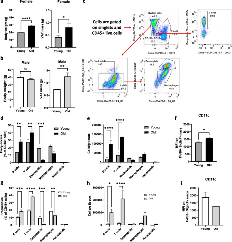
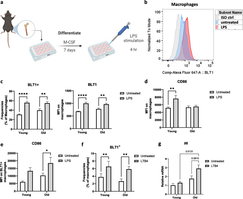
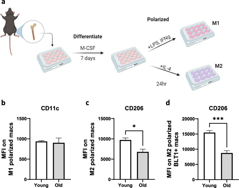
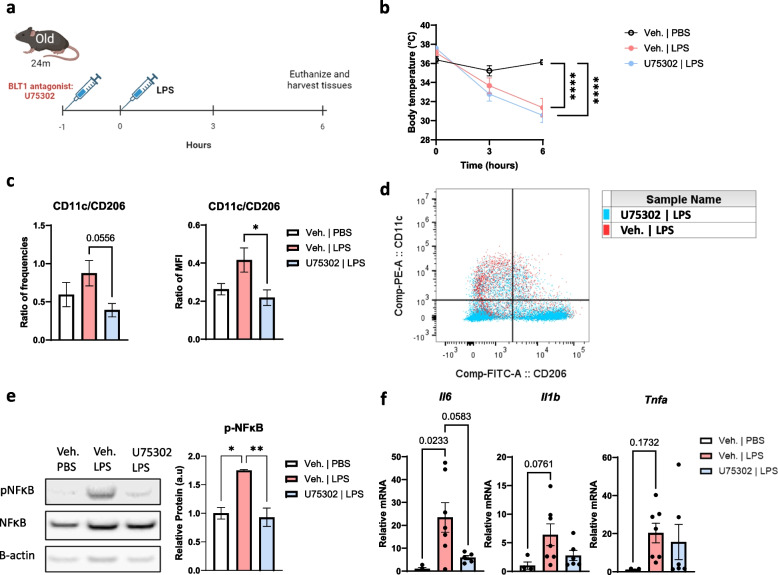
Results: In this study, we investigated BLT1 expression in immune cell subsets within the VAT of aged male and female mice. Moreover, we examined how antagonizing BLT1 signaling could alter the inflammatory response to LPS in aged mice. Our results demonstrate that aged mice exhibit increased adiposity and inflammation, characterized by elevated frequencies of B and T cells, along with pro-inflammatory macrophages in VAT. BLT1 expression is the highest in VAT macrophages. LPS and LTB4 treatment result in increased BLT1 in young and aged bone marrow-derived macrophages (BMDMs). However, LTB4 treatment resulted in amplified Il6 from aged, but not young BMDMs. Treatment of aged mice with the BLT1 antagonist, U75302, followed by LPS-induced endotoxemia resulted in an increase in anti-inflammatory macrophages, reduced phosphorylated NFκB and reduced Il6.

Conclusions: This study provides valuable insights into the age- and sex- specific changes in BLT1 expression on immune cell subsets within VAT. This study offers support for the potential of BLT1 in modulating inflammation in aging.

Keywords: Aging; Inflammation; Leukotriene B4 receptor 1 (BLT1); Macrophage; Sepsis; Visceral adipose tissue.


Conflict of interest statement

The authors declare no competing interests.

Figures

Fig. 1
Fig. 1
Comparison of immune cell population in VAT of young (3–8 months) and aged (20–24 months) mice. a Quantification of young and aged female mice body weight (g) and VAT mass (g). b Quantification of young and aged male mice body weight (g) and VAT mass (g). c Flow cytometry gating strategy of immune cell subsets. d Frequencies of immune cell subsets in female mice. e Cells/g tissue of immune cell subset in female mice. f CD11c MFI of F4/80+ SiglecF macrophages from female mice. g Frequencies of immune cell subsets in male mice. h Cells/g tissue of immune cell subsets in male mice. i CD11c MFI of F4/80+ SiglecF. macrophages of male mice. Female (a, c-f) and male (b, g-i) mice were used in this experiment (n = 4–5/group). Chart error bars represent mean ± SEM. Statistical analysis was conducted using 2-way ANOVA with a post-hoc test of Sidak (d-e, g-h) or non-parametric T-tests (a-b, f, i). *p ≤ 0.05, **p ≤ 0.01, ***p ≤ 0.001, ****p ≤ 0.0001
Fig. 2
Fig. 2
Comparison of BLT1 in immune cell subsets of young (3–8 months) and aged (20–24 months) mice. a Histogram to identify BLT1+ cells. b BLT1 MFI of immune cell subset in VAT, peritoneal exudate cells (PEC), and spleen. c Histogram of BLT1 in VAT immune cell subset. d Frequencies and count of BLT1+ cells in immune cell subset in female young vs. aged mice. e Frequencies and count of BLT1+ cells in immune cell subset in male young vs. aged mice. Female (a-d) and male (e) mice were used in this experiment (n = 4–5/group). Error bars represent mean ± SEM. Statistical analysis was conducted using paired one-way ANOVA with a post-hoc test of Tukey (b) or 2-way ANOVA with a post-hoc test of Sidak (d-e). *p ≤ 0.05, **p ≤ 0.01, ***p ≤ 0.001, ****p ≤ 0.0001
Fig. 3
Fig. 3
Pro-inflammatory (CD86+) phenotype in LPS-treated BLT1+ BMDMs. a Schematic represents the method used to differentiate BMDMs and treat with 1 µg/ml of LPS. b Representative histogram of BLT1 of macrophages in untreated vs. LPS treated BMDMs. c Frequency of BLT1+ cells and BLT1 MFI on macrophages with or without LPS treatment. d CD86 MFI on macrophages with or without LPS treatment. e CD86 MFI on BLT1.+ macrophages with or without LPS treatment. f BLT1 frequencies of macrophages with or without LTB4 treatment. g II6 expression of BMDMs treated with or without LTB4. mRNA expression is normalized to 18 s and the young untreated group. Male mice were used in this experiment (n = 5–6/group). Error bars represent mean ± SEM. Statistical analysis was conducted using 2-way ANOVA with a post-hoc test of Sidak. *p ≤ 0.05, **p ≤ 0.01, ***p ≤ 0.001, ****p ≤ 0.0001
Fig. 4
Fig. 4
Pro-inflammatory (CD11c+) and anti-inflammatory (CD206+) phenotype macrophages in M1 and M2 polarized BMDMs of young and aged mice. a Schematic represents the method used to differentiate and polarize BMDMs. CD11c MFI was examined in M1 polarized BMDMs, and CD206 was examined in M2 polarized BMDMs b CD11c MFI of macrophages in M1 polarized BMDMs. c CD206 MFI of macrophages in M2 polarized BMDMs. d CD206 MFI in BLT1.+ macrophages in M2 polarized BMDMs. Male mice were used in this experiment (n = 4/group). Error bars represent mean ± SEM. Statistical analysis was conducted using non-parametric T-tests. *p ≤ 0.05, **p ≤ 0.01, ***p ≤ 0.001, ****p ≤ 0.0001
Fig. 5
Fig. 5
BLT1 antagonist (U75302) in LPS-exposed aged (24 months) male mice. a Schematic represents the experimental design. Mice were injected with 3 mg/kg LPS and 1 mg/kg U75302 (BLT1 antagonist) through IP. b Body temperature of vehicle, LPS treated and LPS with BLT1 antagonist treated mice. c Ratio of CD11c+ and CD206+ to frequency and MFI of F4/80+ SiglecF. macrophages in VAT. d Dot plot of LPS vs. LPS with BLT1 antagonist treated mouse in VAT. e Protein expression of NF-κB and phospho-NF-κB in VAT of vehicle, LPS treated and LPS with BLT1 antagonist treated mice. Phospho-NF-κB is nomalized to NF-κB and β-actin. f Relative gene expression in VAT of vehicle, LPS treated and LPS with BLT1 antagonist treated mice. mRNA expression is normalized to 18 s and the vehicle group. Male mice were performed in this experiment (n = 4 in vehicle, n = 8 in LPS, n = 7 in LPS with BLT1 antagonist). Error bars represent mean ± SEM. Statistical analysis was conducted using one-way ANOVA with a post-hoc test of Tukey. *p ≤ 0.05, **p ≤ 0.01, ***p ≤ 0.001, ****p ≤ 0.0001

Similar articles

Cited by

References

    1. Huang W, Hickson LJ, Eirin A, Kirkland JL, Lerman LO. Cellular senescence: the good, the bad and the unknown. Nat Rev Nephrol. 2022;18(10):611–27. 10.1038/s41581-022-00601-z - DOI - PMC - PubMed
    1. Frasca D, Romero M, Diaz A, Garcia D, Thaller S, Blomberg BB. B Cells with a Senescent-Associated Secretory Phenotype Accumulate in the Adipose Tissue of Individuals with Obesity. Int J Mol Sci. 2021;22(4). - PMC - PubMed
    1. Pawelec G. Age and immunity: What is “immunosenescence”? Exp Gerontol. 2018;105:4–9. 10.1016/j.exger.2017.10.024 - DOI - PubMed
    1. Dahlquist KJV, Camell CD. Aging Leukocytes and the Inflammatory Microenvironment of the Adipose Tissue. Diabetes. 2022;71(1):23–30. 10.2337/dbi21-0013 - DOI - PMC - PubMed
    1. Frasca D, Blomberg BB. Inflammaging decreases adaptive and innate immune responses in mice and humans. Biogerontology. 2016;17(1):7–19. 10.1007/s10522-015-9578-8 - DOI - PMC - PubMed

LinkOut - more resources