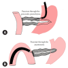
Safety and efficacy of trans-afferent loop endoscopic ultrasound-guided pancreaticojejunostomy for post pancreaticoduodenectomy anastomotic stricture using the forward-viewing echoendoscope: a retrospective study from Japan
- PMID: 39188116
- PMCID: PMC11983134
- DOI: 10.5946/ce.2024.089
Safety and efficacy of trans-afferent loop endoscopic ultrasound-guided pancreaticojejunostomy for post pancreaticoduodenectomy anastomotic stricture using the forward-viewing echoendoscope: a retrospective study from Japan
Abstract
Background/aims: Endoscopic ultrasound (EUS)-guided pancreatic duct drainage is a well-established procedure for managing pancreaticojejunostomy anastomotic strictures (PJAS) post-Whipple surgery. In this study, we examined the effectiveness and safety of EUS-guided pancreaticojejunostomy (EUS-PJS).
Methods: This retrospective, single-arm study was performed at Aichi Cancer Center Hospital on 10 patients who underwent EUS-guided pancreaticojejunostomy through the afferent jejunal loop using a forward-viewing echoendoscope when endoscopic retrograde pancreatography failed. Our primary endpoint was technical success rate, defined as successful stent insertion. The secondary endpoints were early and late adverse events.
Results: A total of 10 patients underwent EUS-PJS between February 2019 and October 2023. The technical success rate was 100%. The median procedure time was 23.5 minutes. No remarkable early or late adverse events related to the procedure, except for fever, occurred in two patients. The median follow-up duration was 9.5 months, and the median number of stent exchanges was two. A stent-free state was achieved in three patients.
Conclusions: EUS-PJS for PJAS management after pancreaticoduodenectomy appears to be an effective and safe procedure with the potential advantages of fewer reinterventions and the creation of a permanent drainage fistula.
Keywords: Drainage; Endosonography; Gastrointestinal endoscopy; Pancreatic ducts; Pancreaticoduodenectomy.
Conflict of interest statement
The authors have no potential conflicts of interest.
Figures


Comment in
-
Transforming endoscopic approaches to post-pancreaticoduodenectomy anastomotic strictures: beyond the surface.Clin Endosc. 2025 Mar;58(2):259-260. doi: 10.5946/ce.2025.004. Epub 2025 Mar 24. Clin Endosc. 2025. PMID: 40200661 Free PMC article. No abstract available.
References
-
- Cioffi JL, McDuffie LA, Roch AM, et al. Pancreaticojejunostomy stricture after pancreatoduodenectomy: outcomes after operative revision. J Gastrointest Surg. 2016;20:293–299. - PubMed
-
- Cotton PB, Eisen GM, Aabakken L, et al. A lexicon for endoscopic adverse events: report of an ASGE workshop. Gastrointest Endosc. 2010;71:446–454. - PubMed
-
- Kikuyama M, Itoi T, Ota Y, et al. Therapeutic endoscopy for stenotic pancreatodigestive tract anastomosis after pancreatoduodenectomy (with videos) Gastrointest Endosc. 2011;73:376–382. - PubMed
LinkOut - more resources
Full Text Sources

