Review
. 2023 Oct 27;2(4):e20230021.
doi: 10.1002/SMMD.20230021. eCollection 2023 Nov.

Emerging biotechnologies and biomedical engineering technologies for hearing reconstruction

Affiliations
Review

Emerging biotechnologies and biomedical engineering technologies for hearing reconstruction

Yangnan Hu et al. Smart Med. .

Abstract

Hearing impairment is a global health problem that affects social communications and the economy. The damage and loss of cochlear hair cells and spiral ganglion neurons (SGNs) as well as the degeneration of neurites of SGNs are the core causes of sensorineural hearing loss. Biotechnologies and biomedical engineering technologies provide new hope for the treatment of auditory diseases, which utilizes biological strategies or tissue engineering methods to achieve drug delivery and the regeneration of cells, tissues, and even organs. Here, the advancements in the applications of biotechnologies (including gene therapy and cochlear organoids) and biomedical engineering technologies (including drug delivery, electrode coating, electrical stimulation and bionic scaffolds) in the field of hearing reconstruction are presented. Moreover, we summarize the challenges and provide a perspective on this field.

Keywords: drug delivery; electrical stimulation; gene therapy; hearing reconstruction; organoid.


Conflict of interest statement

The authors declare that there are no competing interests.

Figures
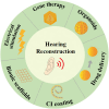
FIGURE 1
FIGURE 1
Schematic illustration of different biotechnologies and biomedical engineering technologies for hearing construction.
FIGURE 2
FIGURE 2
Anatomy of the inner ear in mammals. (A) The location and structure of the inner ear. (B) The cross‐section of the cochlea displaying three fluid‐filled longitudinal tubes. (C) The organ of Corti. Reproduced under terms of the CC‐BY license. Copyright 2020, The Authors, published by MDPI.
FIGURE 3
FIGURE 3
(A) (i) Confocal images showing AAV‐ie targeted SCs in the cochlea. Scale bar: 50 μm. (ii) Statistics of SCs expressing NLS‐mNeonGreen per 100 μm in (i). (iii) Confocal images of different types of SCs in apical turn of cochleae, including HeC, DC, OPC, IPC, and IBC. Scale bar: 20 μm. (B) Confocal projection images showing AAV‐ie‐Atoh1‐induced regeneration of HCs in vivo with stereocilia. Scale bar: 20 μm. Reproduced under terms of the CC‐BY license. Copyright 2019, The Authors, published by Springer Nature. AAV, adeno‐associated virus; DC, Deiters cells; HeC, Hensen's cells; IBC, inner border cells; IPC, inner pillar cells; NLS‐mNeonGreen, nuclear mNeonGreen; OPC, outer pillar cells; SCs, supporting cells.
FIGURE 4
FIGURE 4
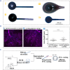
(A) Overview of the inner ear induction method. Reproduced with permission. Copyright 2014, Springer Nature. (B) (i) Immunofluorescence images of cochlear organoids. Myo7a (green) is a marker of HCs, Sox2 (red) is a marker of SCs, and DAPI (blue) represents the nuclei of cells. Scale bar: 50 μm. (ii) Confocal images of co‐localization of CTBP2+ and PSD95+ puncta in cochlear organoids. PSD95 (green) is a postsynaptic marker, CTBP2 (red) is a pre‐synaptic marker, and Atoh1 (blue) represents HCs. Scale bar: 10 μm. Reproduced under terms of the CC‐BY license. Copyright 2022, The Authors, published by John Wiley and Sons. DAPI, 2‐(4‐Amidinophenyl)‐6‐indolecarbamidine dihydrochloride; HeC, Hensen's cells; SCs, supporting cells.
FIGURE 5
FIGURE 5
Drug delivery system for hearing loss treatment. (A) Schematic of ROS‐responsive nanoparticles delivering BBR to the inner ear. Reproduced with permission. Copyright 2021, American Chemical Society. (B) (i) The preparation process of GM@PDA@Lipo‐Ebselen and the diagram of injecting microspheres onto RWM. (ii) Diagram of the process of drugs entering into the inner ear through RWM. Reproduced with permission. Copyright 2022, John Wiley and Sons. (C) (i) Peristaltic micropump for inner ear administration. (ii) Photographs of a mouse after subcutaneous implanting a peristaltic micropump 1 month. Reproduced with permission. Copyright 2019, Elsevier. BBR, berberine; ROS, reactive oxygen species; RWM, round window membrane.
FIGURE 6
FIGURE 6
(A) Bare cochlear electrode (i) and cochlear electrode coated with conductive PEDOT (ii), RGD‐alginate hydrogel and PEDOT (iii), and dehydrated RGD‐alginate hydrogel and PEDOT (iv). Reproduced with permission. Copyright 2012, Elsevier. (B) (i–ii) Immunofluorescence images of SGN explants that were treated with control nanoparticles (i) or nanoparticles carring BDNF (ii), scale bars represent 200 μm in (i–ii). (iii) Axon length of SGN explants treated with different nanoparticles. Reproduced with permission. Copyright 2021, Elsevier. (C) Schematic diagram of the fabrication of DXM/PCL cochlear electrode coatings. Reproduced under terms of the CC‐BY license. Copyright 2021, The Authors, published by Informa UK Limited. BDNF, brain derived neurotrophic factor; DXM, dexamethasone; PCL, poly‐e‐caprolactone; PEDOT, poly (3, 4‐ethylenedioxythiophene); SGN, spiral ganglion neuron.
FIGURE 7
FIGURE 7
(A) Schematic diagram (i) and photograph (ii) of the printed circuit board connected with CI. Reproduced with permission. Copyright 2019, American Chemical Society. (B) Schematic diagram of the fabrication of 3D electrical stimulation culture system based on CI and Ti3C2Tx MXene‐Matrigel hydrogel and its regulation on SGNs. Reproduced with permission. Copyright 2022, The Authors, published by American Chemical Society. (C) Immunofluorescence images of NSCs induced by the lower frequency electric‐acoustic stimulation produced by CI/graphene EAS system. EN 1, 11, 22 referred to high‐, middle‐, and low‐frequency EAS severally. EN22‐10, EN22‐20, and EN22‐40 represented EAS with EN22 for 10, 20, 40 min per day, respectively. Reproduced with permission. Copyright 2021, Royal Society of Chemistry. (D) (i–v) Optical images of Rosenthal's canal at the cochlear location. (vi) The average of SGN densities at different locations. Reproduced with permission. Copyright 2010, Elsevier. CI, cochlear implant; EAS, electric‐acoustic stimulation; EN, electrode number; NSCs, neural stem cells; SGN, spiral ganglion neuron.
FIGURE 8
FIGURE 8
(A) (i) Immunofluorescence images of SGNs on different substrates. Tuj‐1 (red) is a marker of neurons and 9 DAPI (blue) represents the nuclei of cells. (ii) Polar histograms of neurite distribution on different substrates. Reproduced with permission. Copyright 2022, Elsevier. (B) (i) The preparation diagram of the SA‐CNT/GelMA‐modified wings. (ii) Growth and extension of SGNs on the modified wings. Reproduced with permission. Copyright 2021, John Wiley and Sons. DAPI, 2‐(4‐Amidinophenyl)‐6‐indolecarbamidine dihydrochloride; SA‐CNT, super‐aligned carbon‐nanotube sheets; SGN, spiral ganglion neuron.

References

    1. Géléoc G. S., Holt J. R., Science 2014, 344, 1241062. - PMC - PubMed
    1. Liberman M. C., F1000Res. 2017, 6, 927. - PMC - PubMed
    1. Sergeyenko Y., Lall K., Liberman M. C., Kujawa S. G., J. Neurosci. 2013, 33, 13686. - PMC - PubMed
    1. Meena R., Ayub M., J. Ayub Med. Coll. Abbottabad 2017, 29, 671. - PubMed
    1. Li L., Chao T., Brant J., O'Malley B. Jr., Tsourkas A., Li D., Adv. Drug Delivery Rev. 2017, 108, 2. - PMC - PubMed

LinkOut - more resources