

Case Reports
doi: 10.1016/j.radcr.2024.07.019.
eCollection 2024 Oct.
Chronic cough and noisy breathing: An 11-year journey to diagnosis and successful treatment
Affiliations
- PMID: 39188624
- PMCID: PMC11345125
- DOI: 10.1016/j.radcr.2024.07.019
Item in Clipboard
Case Reports
Chronic cough and noisy breathing: An 11-year journey to diagnosis and successful treatment
Radiol Case Rep.
.
Abstract
Foreign body aspiration in an uncommon entity in adults which often leads to delays in diagnosis. Adults with long-standing foreign bodies in the airway can result in various complications including bronchiectasis, atelectasis and lung fibrosis. We describe the case of a primary school teacher who was diagnosed with foreign body aspiration 11 years after the aspiration event. Delays in diagnosis led to her receiving multiple doses of antibiotics including a course of antituberculous therapy.
Keywords: Bronchoscopy; Chronic cough; Foreign body.
© 2024 The Authors. Published by Elsevier Inc. on behalf of University of Washington.
Figures

Fibrotic changes and scattered cavitation seen in the left apical region, reduced left lung volume with traction bronchiectasis. There were also small centrilobular nodules in a linear branching pattern in the left lung resembling tree in bud appearance suggestive for bronchiolitis.
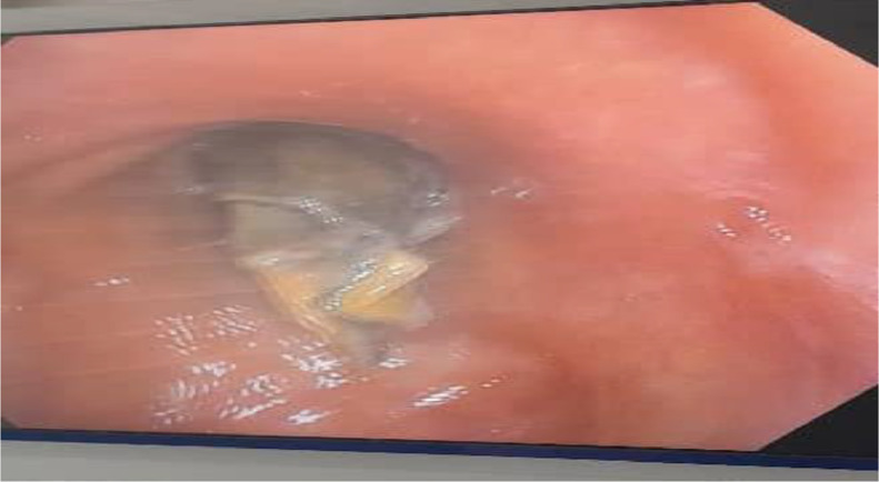
Bronchoscopy image showing a foreign body in the left main bronchus.

Piece of sweet shell (plastic wrap) removed from the left main bronchus.

Chest-Xray after foreign body removal showing fibrosis, bronchiectasis and reduced left lung volume with few small bullae in upper pole.

Review of the first CT scan images showed a high-density foreign body (approximately – 250 HU density) is seen in the left main bronchus, approximately about 2.86 cm distal to carina impacted at the bifurcation into the 2 lobar bronchi, the left upper and lower lobar bronchi. (black arrow). Furthermore, there was a hypodense opacity in the left lower lobe that was best seen in coronal views with classic finger in glove appearance which represents mucous filling dilated bronchi (20-28 HU density) consistent with mucoid impaction.
References
Publication types
LinkOut - more resources
Full Text Sources
