. 2024 Aug 12:18:1415134.
doi: 10.3389/fnins.2024.1415134. eCollection 2024.

Altered brain function during movement programming is linked with motor deficits after stroke: a high temporal resolution study

Affiliations

Altered brain function during movement programming is linked with motor deficits after stroke: a high temporal resolution study

Célia Delcamp et al. Front Neurosci. .

Abstract

Introduction: Stroke leads to motor deficits, requiring rehabilitation therapy that targets mechanisms underlying movement generation. Cortical activity during the planning and execution of motor tasks can be studied using EEG, particularly via the Event Related Desynchronization (ERD). ERD is altered by stroke in a manner that varies with extent of motor deficits. Despite this consensus in the literature, defining precisely the temporality of these alterations during movement preparation and performance may be helpful to better understand motor system pathophysiology and might also inform development of novel therapies that benefit from temporal resolution.

Methods: Patients with chronic hemiparetic post-stroke (n = 27; age 59 ± 14 years) and age-matched healthy right-handed control subjects (n = 23; 59 ± 12 years) were included. They performed a shoulder rotation task following the onset of a stimulus. Cortical activity was recorded using a 256-electrode EEG cap. ERD was calculated in the beta frequency band (15-30 Hz) in ipsilesional sensorimotor cortex, contralateral to movement. The ERD was compared over time between stroke and control subjects using permutation tests. The correlation between upper extremity motor deficits (assessed by the Fugl-Meyer scale) and ERD over time was studied in stroke patients using Spearman and permutation tests.

Results: Patients with stroke showed on average less beta ERD amplitude than control subjects in the time window of -350 to 50 ms relative to movement onset (t(46) = 2.8, p = 0.007, Cohen's d = 0.31, 95% CI [0.22: 1.40]). Beta-ERD values correlated negatively with the Fugl-Meyer score during the time window -200 to 400 ms relative to movement onset (Spearman's r = -0.54, p = 0.003, 95% CI [-0.77 to -0.18]).

Discussion: Our results provide new insights into the precise temporal changes of ERD after hemiparetic stroke and the associations they have with motor deficits. After stroke, the average amplitude of cortical activity is reduced as compared to age-matched controls, and the extent of this decrease is correlated with the severity of motor deficits; both were true during motor programming and during motor performance. Understanding how stroke affects the temporal dynamics of cortical preparation and execution of movement paves the way for more precise restorative therapies. Studying the temporal dynamics of the EEG also strengthens the promising interest of ERD as a biomarker of post-stroke motor function.

Keywords: biomarker; cortical activity; event-related desynchronization; motor control; premovement.


Conflict of interest statement

The authors declare that the research was conducted in the absence of any commercial or financial relationships that could be construed as a potential conflict of interest.

Figures

Figure 1
Figure 1
(A) Rest position of subjects with forearm in the tabletop splint. (B) Enlargement of the red box from (A) (with contrast adjusted for visibility), with the green arrow pointing to the 2 mm gap between splint and switch. This allows movement of the forearm in the plane of the table, after which the splint contacts the switch and can be displaced no further. In this image, the subject’s forearm is on the right side of the image and a switch is on the left side. (C) Representation of study design composed of four blocks of 20 movements. Each movement starts with rest, followed by movement cue, followed by return of the arm to the basal position, as outlined in vertically ascending order.
Figure 2
Figure 2
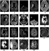
Representative images of the infarct are shown for patients with stroke. The arrow indicates the lesion. Images could not be retrieved from outside medical records in eight patients. L, left; R, right.
Figure 3
Figure 3
Temporal evolution of beta-ERD (blue line = stroke patients; red line = healthy controls) in C3 and surrounding leads. The shaded area represents the time window where the two subject groups show a statistically significant difference in amplitude, which occurred both before and during movement. The dotted black line indicates the start of movement; the solid black line, the average appearance of the stimulus.
Figure 4
Figure 4
Topographical representation of the beta-ERD for the time window from −350 to +50 ms, which in (A) stroke patients shows a similar but smaller spatial distribution as compared to (B) healthy controls. White dots indicate C3 and the six leads surrounding it, which for patients with stroke is the ipsilesional hemisphere and for healthy controls it is the left hemisphere.
Figure 5
Figure 5
Individual and boxplot representation of the beta-ERD for the time window from −350 to +50 ms. In each box, the center line represents the median, the top, and bottom of the box correspond to the 25th and 75th percentiles, and the whiskers represent extreme values excluding outliers (<25% −1.5 interquartile range and > 75% +1.5 interquartile range). The amplitude of the beta-ERD was decreased in patients with stroke compared to age-matched healthy controls (Mann–Whitney U-test: p = 0.02, Rank-Biserial correlation = 0.39, 95% CI [0.09: 0.63]).
Figure 6
Figure 6
Temporal evolution of the correlation between beta-ERD and the upper extremity Fugl-Meyer score; gray shaded area represents a random distribution derived from permutation analysis, with values outside the gray box indicating a significant (p < 0.05) correlation. The dotted black line represents the start of movement; the solid black line, the average appearance of the stimulus. Negative values for ρ indicate that a more negative ERD correlates with higher UE-FM score—more cortical activity (a more negative ERD) is associated with better motor status (higher UE-FM score).
Figure 7
Figure 7
Correlation between beta-ERD for the time window from −200 to +400 ms and the upper extremity Fugl-Meyer score (Spearman’s r = −0.54, 95% CI [−0.77 to −0.18], p = 0.003).

References

    1. Bartur G., Pratt H., Soroker N. (2019). Changes in mu and beta amplitude of the EEG during upper limb movement correlate with motor impairment and structural damage in subacute stroke. Clin. Neurophysiol. 130, 1644–1651. doi: 10.1016/j.clinph.2019.06.008, PMID: - DOI - PubMed
    1. Belkacem A. N., Jamil N., Khalid S., Alnajjar F. (2023). On closed-loop brain stimulation systems for improving the quality of life of patients with neurological disorders. Front. Hum. Neurosci. 17:1085173. doi: 10.3389/fnhum.2023.1085173, PMID: - DOI - PMC - PubMed
    1. Bigot J., Longcamp M., Dal Maso F., Amarantini D. (2011). A new statistical test based on the wavelet cross-Spectrum to detect time-frequency dependence between non-stationary signals: application to the analysis of Cortico-muscular interactions. NeuroImage 55, 1504–1518. doi: 10.1016/j.neuroimage.2011.01.033 - DOI - PubMed
    1. Chalard A., Amarantini D., Tisseyre J., Marque P., Gasq D. (2020). Spastic co-contraction is directly associated with altered cortical Beta oscillations after stroke. Clin. Neurophysiol. 131, 1345–1353. doi: 10.1016/j.clinph.2020.02.023, PMID: - DOI - PubMed
    1. Delcamp C., Gasq D., Cormier C., Amarantini D. (2023). Corticomuscular and intermuscular coherence are correlated after stroke: a simplified motor control? Brain Commun. 5:fcad187. doi: 10.1093/braincomms/fcad187, PMID: - DOI - PMC - PubMed