Review
. 2024 Aug 22:20:2084-2107.
doi: 10.3762/bjoc.20.180. eCollection 2024.

Computational toolbox for the analysis of protein-glycan interactions

Affiliations
Review

Computational toolbox for the analysis of protein-glycan interactions

Ferran Nieto-Fabregat et al. Beilstein J Org Chem. .

Abstract

Protein-glycan interactions play pivotal roles in numerous biological processes, ranging from cellular recognition to immune response modulation. Understanding the intricate details of these interactions is crucial for deciphering the molecular mechanisms underlying various physiological and pathological conditions. Computational techniques have emerged as powerful tools that can help in drawing, building and visualising complex biomolecules and provide insights into their dynamic behaviour at atomic and molecular levels. This review provides an overview of the main computational tools useful for studying biomolecular systems, particularly glycans, both in free state and in complex with proteins, also with reference to the principles, methodologies, and applications of all-atom molecular dynamics simulations. Herein, we focused on the programs that are generally employed for preparing protein and glycan input files to execute molecular dynamics simulations and analyse the corresponding results. The presented computational toolbox represents a valuable resource for researchers studying protein-glycan interactions and incorporates advanced computational methods for building, visualising and predicting protein/glycan structures, modelling protein-ligand complexes, and analyse MD outcomes. Moreover, selected case studies have been reported to highlight the importance of computational tools in studying protein-glycan systems, revealing the capability of these tools to provide valuable insights into the binding kinetics, energetics, and structural determinants that govern specific molecular interactions.

Keywords: MD; computational tools; glycan–protein interactions; molecular recognition.


Figures

Figure 1
Figure 1
Carbohydrate conformational variability. a) Illustration of Φ, Ψ and ω dihedral angles for a representative trisaccharide coloured according to the symbol nomenclature for glycans (SNFG). b) On the left: Pseudo-rotational wheel depiction of five-membered ring structures showcasing envelope (E) and twist (T) conformations. On the right: Glove representation illustrating the puckering of six-membered pyranoside ring conformations. c) Equilibrium between the low-energy solution conformations of the iduronic acid. The exclusive (Nuclear Overhauser Effect) 1H–1H NOE contacts characteristic of each conformation, 1C4, 4C1 and 2S0, are also depicted.
Figure 2
Figure 2
Monosaccharides diversity in eukaryotes and bacteria. a) Eukaryotic monosaccharides. b) Examples of some peculiar bacterial monosaccharides, including hexuronic acids, heptoses, or octulosonic acids. The SNFG symbol [11] of each monosaccharide is also reported (if any).
Figure 3
Figure 3
Different glycan representations. The 3’-sialyllactosamine is depicted according to the a) IUPAC nomenclature b) chemical representation as implemented in ChemDraw suite [46] c) GlycoCT nomenclature d) SNFG and 3D SNFG nomenclature e) VMD 3D-SNFG plugin f) MolStar representation.
Figure 4
Figure 4
Visualisation programs. Different representation of a protein–ligand complex by using the most used visualisation programs reported in this review. The previously published complex [155] between the Ca2+ dependant C-type lectin, DC-SIGN (PDB: 1SL4) [156], and a tetrasaccharide composed of mannose and rhamnose residues, has been used to highlight the main advantages of each visualisation program applied to protein–glycan complexes.
Figure 5
Figure 5
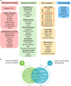
A schematic representation of useful computational methods to study protein–glycan interactions. a) Workflow including different key steps needed to analyse protein–glycan interactions: 1. Building/choosing the appropriate glycan/protein 3D structure; 2. Modelling protein–glycan complex; 3. Running and analysing MD simulations; 4. Processing and visualising the results. b) Summary of the presented tools for building structural model and/or generating topology files of glycan/protein structures.

References

    1. Kuo J C-H, Paszek M J. Annu Rev Cell Dev Biol. 2021;37:257–283. doi: 10.1146/annurev-cellbio-120219-054401. - DOI - PMC - PubMed
    1. Flynn R A, Pedram K, Malaker S A, Batista P J, Smith B A H, Johnson A G, George B M, Majzoub K, Villalta P W, Carette J E, et al. Cell. 2021;184(12):3109–3124.e22. doi: 10.1016/j.cell.2021.04.023. - DOI - PMC - PubMed
    1. Varki A. Glycobiology. 2017;27:3–49. doi: 10.1093/glycob/cww086. - DOI - PMC - PubMed
    1. Varki A, Kornfeld S. In: Cummings R D, Esko J D, Stanley P, et al., editors. New York, NY, USA: Cold Spring Harbor; 2022. pp. 61–91.
    1. Hart G W, Copeland R J. Cell. 2010;143:672–676. doi: 10.1016/j.cell.2010.11.008. - DOI - PMC - PubMed

LinkOut - more resources