. 2024 Aug 9;13(3):68.
doi: 10.3390/antib13030068.

Novel Monoclonal Antibody Specific toward Amyloid-β Binds to a Unique Epitope within the N-Terminal Region

Affiliations

Novel Monoclonal Antibody Specific toward Amyloid-β Binds to a Unique Epitope within the N-Terminal Region

Giavanna Paterno et al. Antibodies (Basel). .

Abstract

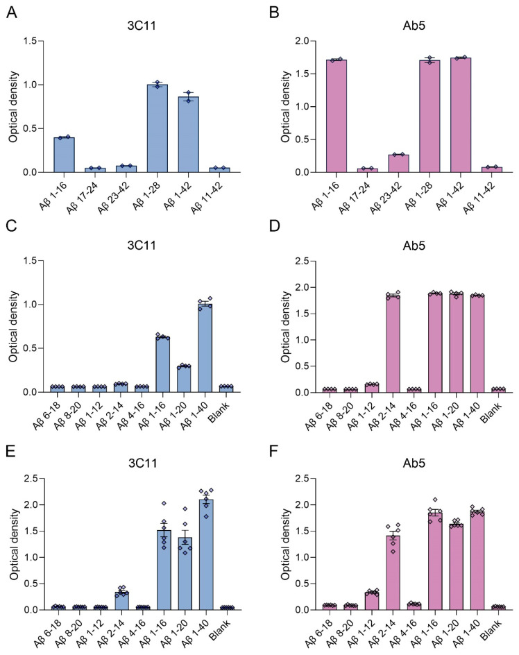
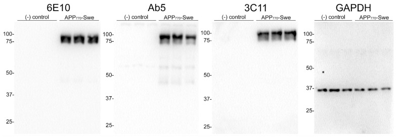
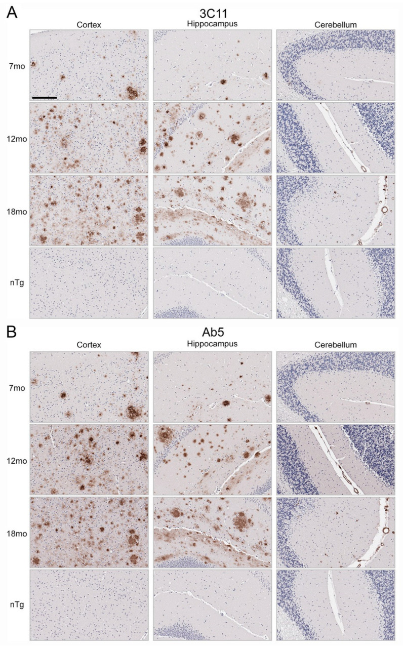
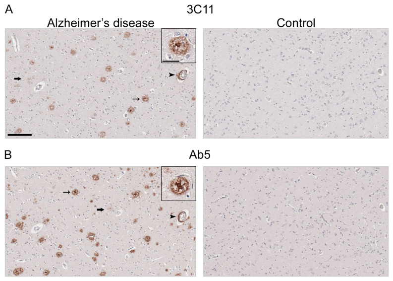
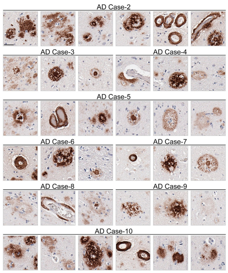
Amyloid-β (Aβ) deposition throughout the neuroaxis is a classical hallmark of several neurodegenerative diseases, most notably Alzheimer's disease (AD). Aβ peptides of varied length and diverse structural conformations are deposited within the parenchyma and vasculature in the brains of individuals with AD. Neuropathologically, Aβ pathology can be assessed using antibodies to label and characterize their features, which in turn leads to a more extensive understanding of the pathological process. In the present study, we generated a novel monoclonal antibody, which we found to be specific for the N-terminal region of Aβ. This antibody reacted to amyloid precursor protein expressed in cultured cells and labels Aβ plaques and cerebral amyloid angiopathy in brain tissue from a mouse model of amyloidosis as well as post-mortem brain tissue from patients diagnosed with AD. This highly specific novel antibody will serve as a unique tool for future studies investigating Aβ deposition in novel mouse models and cross-sectional studies using post-mortem human tissue.

Keywords: Alzheimer’s disease; Amyloid-β; antibody; pathology; plaques.


Conflict of interest statement

The authors declare no conflict of interest.

Figures

Figure 1
Figure 1
ELISA characterization of the novel mouse monoclonal antibody 3C11 using synthetic Aβ peptides. ELISA using method 1 (AD) or 2 (E,F) was performed as described in Section 2. Plates were coated with synthetic Aβ peptides shown in Table 1 or uncoated (Blank) followed by incubation with 3C11 (A,C,E) or Ab5 (B,D,F). 3C11 immunoreactivity was compared to antibody Ab5. Data are shown as mean +/− SEM.
Figure 2
Figure 2
Immunoblot analysis demonstrating specificity of 3C11 monoclonal antibody. HEK293T cells were transfected in triplicate using plasmids expressing Amyloid Precursor Protein (APP) isoform 770 with familial AD Swedish mutation (K670N/M671L) (APP770-Swe) or empty vector (-) control. Cell harvest and western blotting were performed as described in Section 2. Antibodies 6E10 and Ab5 were used as controls for APP and GAPDH as a loading control. The mobility of molecular mass markers is indicated on the left side of each immunoblot.
Figure 3
Figure 3
Immunohistochemical staining of Aβ deposits in brain tissue of TgCRND8 mouse model of amyloidosis. Immunohistochemical staining using monoclonal antibodies 3C11 (A) and Ab5 (B) of 7-, 12-, and 18-month-old TgCRND8 and 7-month-old nTg mice as described in Section 2. The scale bar represents 150 µm.
Figure 4
Figure 4
Immunohistochemical staining of Alzheimer’s disease human brain tissue using antibody 3C11. Immunohistochemical staining of AD and control mid-frontal cortical tissue using antibodies 3C11 (A) and Ab5 (B) was performed as described in Section 2. Immunohistochemical staining of cases AD-1 and Control-1 are shown. Demographics of cases are shown in Table 2. Thin arrows indicate 3C11 or Ab5 immunoreactive core plaques, thick arrows indicate diffuse plaques, and arrowheads indicate cerebral amyloid angiopathy. Scale bars of low magnification images represent 150 µm, and the inset is 35 µm.
Figure 5
Figure 5
Appreciable diversity of Aβ pathology detected by monoclonal antibody 3C11. Immunohistochemical staining of Aβ pathology using monoclonal antibody 3C11 in Alzheimer’s disease cases is shown. Demographics of cases are shown in Table 2. The scale bar represents 35 µm.

Similar articles

References

    1. McKhann G.M., Knopman D.S., Chertkow H., Hyman B.T., Jack C.R., Kawas C.H., Klunk W.E., Koroshetz W.J., Manly J.J., Mayeux R., et al. The Diagnosis of Dementia Due to Alzheimer’s Disease: Recommendations from the National Institute on Aging-Alzheimer’s Association Workgroups on Diagnostic Guidelines for Alzheimer’s Disease. Alzheimers Dement. 2011;7:263–269. doi: 10.1016/j.jalz.2011.03.005. - DOI - PMC - PubMed
    1. Thal D.R., Rüb U., Orantes M., Braak H. Phases of A Beta-Deposition in the Human Brain and Its Relevance for the Development of AD. Neurology. 2002;58:1791–1800. doi: 10.1212/WNL.58.12.1791. - DOI - PubMed
    1. Braak H., Braak E. Neuropathological Stageing of Alzheimer-Related Changes. Acta Neuropathol. 1991;82:239–259. doi: 10.1007/BF00308809. - DOI - PubMed
    1. Braak H., Alafuzoff I., Arzberger T., Kretzschmar H., Del Tredici K., Alafuzov I., Arzberger T., Kretzschmar H., Del Tredici K. Staging of Alzheimer Disease-Associated Neurofibrillary Pathology Using Paraffin Sections and Immunocytochemistry. Acta Neuropathol. 2006;112:389–404. doi: 10.1007/s00401-006-0127-z. - DOI - PMC - PubMed
    1. DeTure M.A., Dickson D.W. The Neuropathological Diagnosis of Alzheimer’s Disease. Mol. Neurodegener. 2019;14:32. doi: 10.1186/s13024-019-0333-5. - DOI - PMC - PubMed

LinkOut - more resources