Multicenter Study
. 2024 Nov 4;147(11):3714-3726.
doi: 10.1093/brain/awae210.

Neurophysiological markers of motor compensatory mechanisms in early Parkinson's disease

Affiliations
Multicenter Study

Neurophysiological markers of motor compensatory mechanisms in early Parkinson's disease

Massimiliano Passaretti et al. Brain. .

Abstract

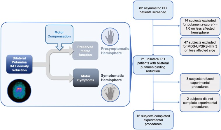
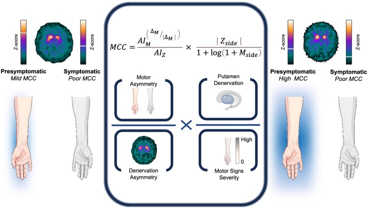
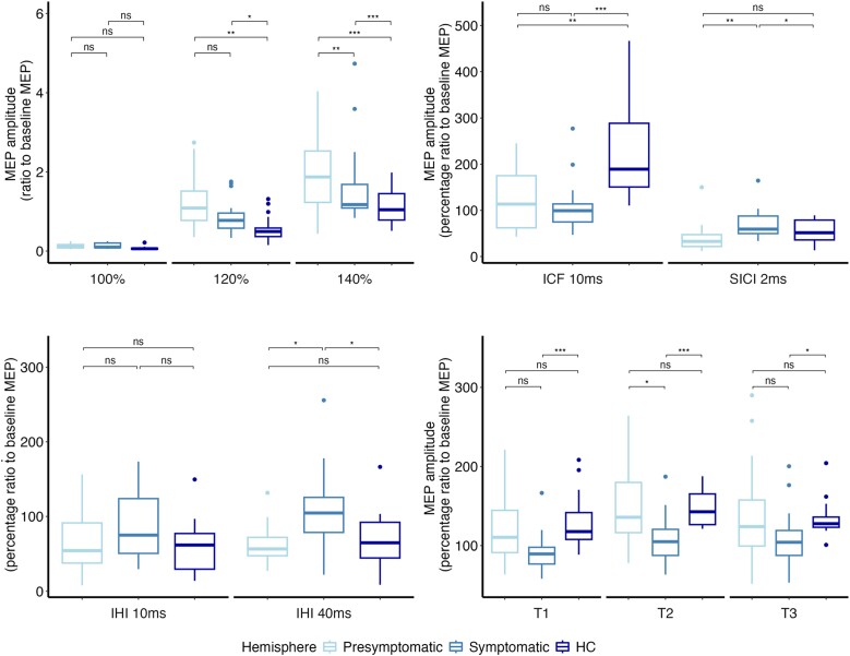
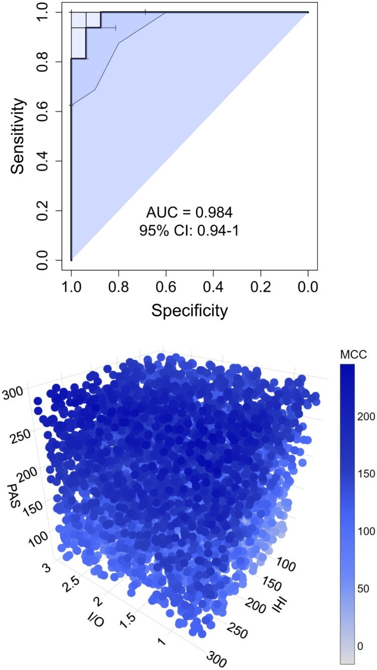
Compensatory mechanisms in Parkinson's disease are defined as the changes that the brain uses to adapt to neurodegeneration and progressive dopamine reduction. Motor compensation in early Parkinson's disease could, in part, be responsible for a unilateral onset of clinical motor signs despite the presence of bilateral nigrostriatal degeneration. Although several mechanisms have been proposed for compensatory adaptations in Parkinson's disease, the underlying pathophysiology is unclear. Here, we investigate motor compensation in Parkinson's disease by investigating the relationship between clinical signs, dopamine transporter imaging data and neurophysiological measures of the primary motor cortex (M1), using transcranial magnetic stimulation in presymptomatic and symptomatic hemispheres of patients. In this cross-sectional, multicentre study, we screened 82 individuals with Parkinson's disease. Patients were evaluated clinically in their medication OFF state using standardized scales. Sixteen Parkinson's disease patients with bilateral dopamine transporter deficit in the putamina but unilateral symptoms were included. Twenty-eight sex- and age-matched healthy controls were also investigated. In all participants, we tested cortical excitability using single- and paired-pulse techniques, interhemispheric inhibition and cortical plasticity with paired associative stimulation. Data were analysed with ANOVAs, multiple linear regression and logistic regression models. Individual coefficients of motor compensation were defined in patients based on clinical and imaging data, i.e. the motor compensation coefficient. The motor compensation coefficient includes an asymmetry score to balance motor and dopamine transporter data between the two hemispheres, in addition to a hemispheric ratio accounting for the relative mismatch between the magnitude of motor signs and dopaminergic deficit. In patients, corticospinal excitability and plasticity were higher in the presymptomatic compared with the symptomatic M1. Also, interhemispheric inhibition from the presymptomatic to the symptomatic M1 was reduced. Lower putamen binding was associated with higher plasticity and reduced interhemispheric inhibition in the presymptomatic hemisphere. The motor compensation coefficient distinguished the presymptomatic from the symptomatic hemisphere. Finally, in the presymptomatic hemisphere, a higher motor compensation coefficient was associated with lower corticospinal excitability and interhemispheric inhibition and with higher plasticity. In conclusion, the present study suggests that motor compensation involves M1-striatal networks and intercortical connections becoming more effective with progressive loss of dopaminergic terminals in the putamen. The balance between these motor networks seems to be driven by cortical plasticity.

Keywords: Parkinson’s disease; cortical plasticity; dopamine transporter compensatory mechanisms.


Conflict of interest statement

All authors declare no competing interests. R.C. has received speaking honoraria from Zambon Italia; Zambon SAU; Bial Italia Srl; Advisory board fees from Bial; Research support from the Ministero Italiano della Salute; Editor-in-Chief of the neuromuscular and movement disorders section of Brain Sciences; Associate Editor of Parkinsonism and Related Disorders and Frontiers in Neurology. T.v.E. is a civil servant of North Rhine-Westphalia (Germany) and an employee of the University Hospital of Cologne, Germany. In the past 2 years, he received honoraria, stipends or speaker fees from the Lundbeck Foundation, Gain Therapeutics, Orion Pharma, Lundbeck Pharma, Atheneum and the International Movement Disorders Society; he receives materials from Life Molecular Imaging and Lilly Pharma; and he owns stocks in the NVIDIA, Microsoft and IBM corporations. Multiple unrelated research projects are currently supported by the German Research Foundation.

Figures

Figure 1
Figure 1
Screening and selection of Parkinson’s disease patients. The initial cohort of 82 subjects had already been screened for other clinical exclusion criteria. DAT = dopamine transporter; MDS-UPDRS = Movement Disorder Society-sponsored revision of the Unified Parkinson’s Disease Rating Scale; PD = Parkinson’s disease.
Figure 2
Figure 2
Graphical representation of motor compensatory coefficient. The MCC is composed of an asymmetry score (left side of the formula) to balance motor and DAT measures between the two hemispheres, and a hemispheric ratio (right side of the formula) accounting for the relative mismatch between the magnitude of motor signs and dopaminergic deficit. Coloured bars generally applied for individual DAT tracer binding are used for graphical purposes only to represent z-scores. Left: Example case of mild motor compensation on the presymptomatic side, with high putamen binding asymmetry, attributable to higher putamen binding in the presymptomatic hemisphere. Right: Example case of high motor compensation, with lower putamen asymmetry, higher clinical asymmetry, lower presymptomatic putamen binding, but still no motor symptoms on the presymptomatic body side. AIM = motor asymmetry index; AIZ = putamen z-score asymmetry index; DAT = dopamine transporter; Mside = motor score of reference side; MCC = motor compensatory coefficient; Zside = putamen z-score of reference side; ΔM = difference of motor scores.
Figure 3
Figure 3
Neurophysiological measures. In each panel are depicted neurophysiological data of symptomatic (light blue) and presymptomatic (intermediate blue) hemispheres of patients with PD and of healthy controls (HCs) (dark blue). Boxes represent the median value and the interquartile interval; lower and upper extremes are settled at 1.5 interquartile intervals under the first and over the third quartile, respectively. Dots are single data-points exceeding the upper and lower limits. The significance of post hoc analyses is represented by bars indicating the comparison, and asterisks indicate the level of significance (*P < 0.05, **P < 0.01 and ***P < 0.001). (A) Input–output curve of MEPs at baseline. The y-axis shows the MEP amplitudes (in millivolts); the x-axis shows the three stimulation intensities (100%, 120% and 140% of resting motor threshold). (B) ICF and SICI at baseline. The y-axis shows the percentage ratio between conditioned and unconditioned MEP amplitudes; the x-axis shows the ISIs. (C) Interhemispheric inhibition at baseline. The y-axis shows the percentage ratio between conditioned and unconditioned MEP amplitudes; the x-axis shows the two ISIs (D) Effect of PAS on corticospinal excitability. The y-axis shows the percentage ratio between conditioned and unconditioned MEP amplitudes. The x-axis shows T1 = 5 min post PAS; T2 = 15 min post PAS; T3 = 30 min post PAS. ICF = intracortical facilitation; IHI = interhemispheric inhibition; ISIs = interstimulus intervals; MEP = motor evoked potential; PAS = paired associative stimulation; PD = Parkinson’s disease; SICI = short-interval intracortical inhibition.
Figure 4
Figure 4
Motor compensation coefficient. The top panel represents the area under the receiver operating characteristic curve of the motor compensation coefficient. The bottom panel represents a three-dimensional scatterplot of linear regression between neurophysiological parameters (I/O = input–output curve; IHI = interhemispheric inhibition; PAS = cortical associative plasticity) and the values of the motor compensation coefficient (MCC) (blue bar) in the presymptomatic hemisphere. AUC = area under the curve; CI = confidence interval.
Figure 5
Figure 5
Graphical representation of major findings of the study. Goal-directed and habitual motor network modifications respond to the reduction in striatal dopamine binding. The cortical neurophysiological response was associated with different striatal binding conditions in the presymptomatic and symptomatic hemispheres, as demonstrated by linear models. In the presymptomatic hemisphere, higher putamen binding was associated with higher M1 corticospinal excitability (habitual putamen–M1 network, enlightened corticoputaminal projections in grey). The reduction in putamen binding in the presymptomatic hemisphere is associated with higher M1 cortical plasticity (goal-directed motor network). In these circumstances, the caudate nucleus exhibits a heightened level of activity compared with the putamen (light blue caudate and corticocaudate networks), and this dominance is correlated with increased levels of associative plasticity (light blue areas and M1–motor associative area networks) and reduced interhemispheric inhibition (dotted blue line between bilateral M1s). The symptomatic hemisphere demonstrates a reduction of cortical plasticity associated with lower caudate binding and a higher putamen/caudate ratio, in keeping with reduced activity of the goal-directed motor network. IHI = interhemispheric inhibition; I/O = input–output curve; M1 = primary motor cortex; PAS = cortical associative plasticity.

Comment in

References

    1. Diederich NJ, Surmeier DJ, Uchihara T, Grillner S, Goetz CG. Parkinson’s disease: Is it a consequence of human brain evolution? Mov Disord. 2019;34:453–459. - PMC - PubMed
    1. Foffani G, Obeso JA. A cortical pathogenic theory of Parkinson’s disease. Neuron. 2018;99:1116–1128. - PubMed
    1. Nambu A. Somatotopic organization of the primate basal ganglia. Front Neuroanat. 2011;5:26. - PMC - PubMed
    1. Kojovic M, Bologna M, Kassavetis P, et al. Functional reorganization of sensorimotor cortex in early Parkinson disease. Neurology. 2012;78:1441–1448. - PMC - PubMed
    1. Bologna M, Paparella G, Fasano A, Hallett M, Berardelli A. Evolving concepts on bradykinesia. Brain. 2020;143:727–750. - PMC - PubMed

Publication types

MeSH terms

Substances