Review
. 2024 Sep 23;52(17):10040-10067.
doi: 10.1093/nar/gkae736.

CRISPR-Cas target recognition for sensing viral and cancer biomarkers

Affiliations
Review

CRISPR-Cas target recognition for sensing viral and cancer biomarkers

Shadi Rahimi et al. Nucleic Acids Res. .

Abstract

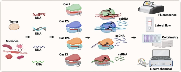
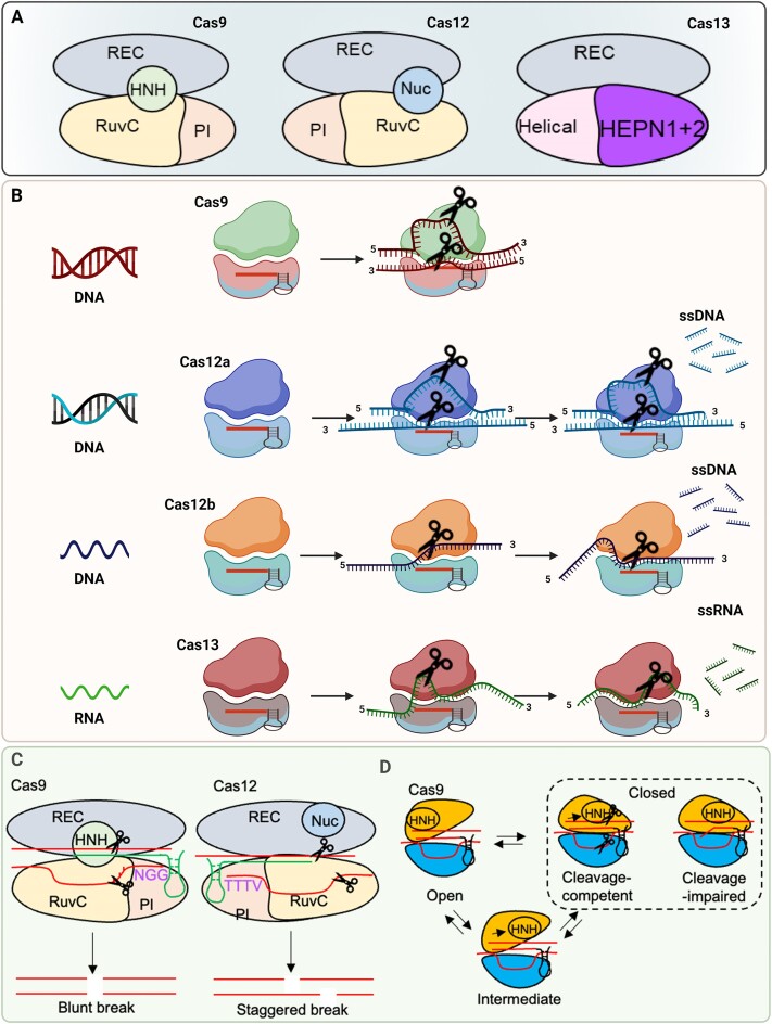
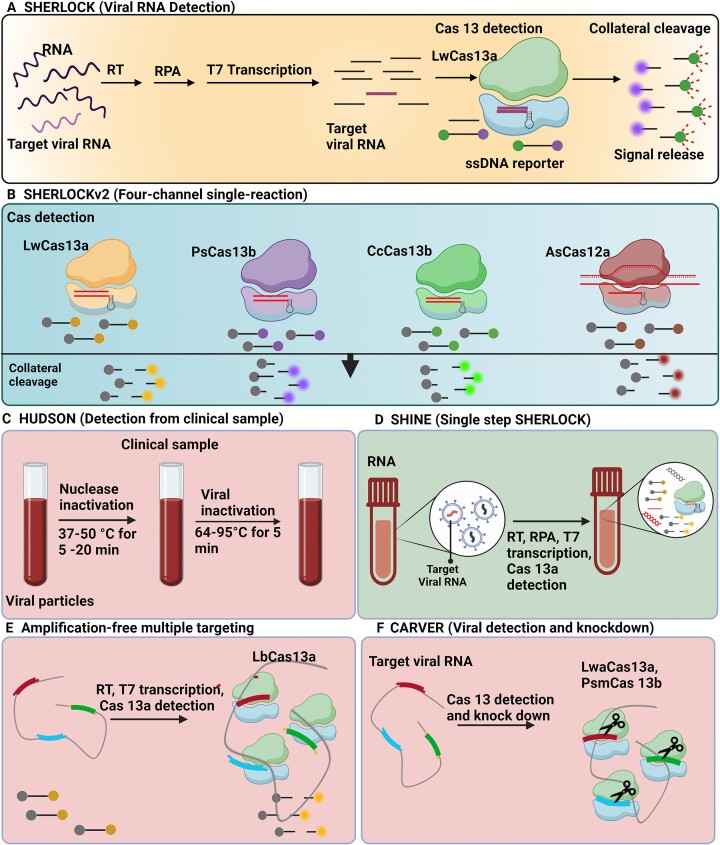
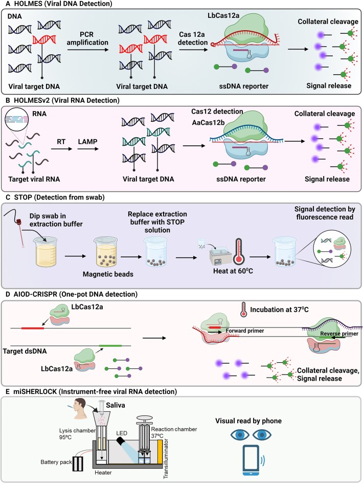
Nucleic acid-based diagnostics is a promising venue for detection of pathogens causing infectious diseases and mutations related to cancer. However, this type of diagnostics still faces certain challenges, and there is a need for more robust, simple and cost-effective methods. Clustered regularly interspaced short palindromic repeats (CRISPRs), the adaptive immune systems present in the prokaryotes, has recently been developed for specific detection of nucleic acids. In this review, structural and functional differences of CRISPR-Cas proteins Cas9, Cas12 and Cas13 are outlined. Thereafter, recent reports about applications of these Cas proteins for detection of viral genomes and cancer biomarkers are discussed. Further, we highlight the challenges associated with using these technologies to replace the current diagnostic approaches and outline the points that need to be considered for designing an ideal Cas-based detection system for nucleic acids.


Figures

Graphical Abstract
Graphical Abstract
Figure 1.
Figure 1.
(A) The structure of Cas9, Cas12 and Cas13 proteins. Cas9 comprises RuvC and HNH nuclease domains, Cas12 comprises single RuvC nuclease domain, and Cas13 contains two conserved HEPN (higher eukaryotes and prokaryotes nucleotide-binding) domains that cleaves RNA. (B) Mechanism of function of Cas9, Cas12 and Cas13 proteins on DNA and RNA. While Cas9 and Cas12 enzymes target DNA, Cas13 cleaves single-stranded RNA targets. Cleavage of surrounding nontargeted nucleic acids after recognition of target is called collateral cleavage activity. Collateral cleavage activity of Cas12 is against ssDNAs while that of Cas13 is against ssRNAs. (C) Cas9 and Cas12 cleavage of target. For Cas9, the PAM sequence (5′-NGG-3′) is located downstream of the spacer on the non-template strand and recognized by the PI domain. The HNH and RuvC domains in Cas9 cleave template and non-template at 3 base pairs upstream of the PAM, respectively, thereby generating a blunt double strand break (DSB). For Cas12, the PAM, typically 5′-TTTV-3′, is located upstream of the spacer and a single nuclease RuvC cut DNA strands in the same nuclease site, generating the staggered double strand break (DSB). (D) Cas9 fluctuates among three major conformational states including open, intermediate, and closed states, that reflects conformational mobility of the catalytic HNH domain.
Figure 2.
Figure 2.
Cas13-based detection methods for viral RNA. (A) SHERLOCK is based on the conversion of RNA to DNA by reverse transcription (RT), recombinase polymerase amplification (RPA)-based amplification, and transcription back to RNA for detection by Cas13, and further Cas13-mediated collateral cleavage of a reporter RNA (38). (B) SHERLOCKv2 in-sample four-channel multiplexing with orthogonal Cas13 and Cas12a enzymes (39). (C) Viral detection directly from bodily fluids using HUDSON (40). (D) RPA-based amplification and Cas13 in a single step using SHINE (42). (E) Amplification-free multiple crRNAs targeting of Cas13 (46). (F) Cas13 for viral detection and knockdown (43).
Figure 3.
Figure 3.
Cas12-based detection methods for viral DNA. (A) Detection of viral target DNA using HOLMES (59). (B) Detection of viral target RNA using HOLMESv2 (17). (C) Simplified viral RNA extraction, isothermal amplification, and Cas12b detection using STOP at a single temperature (60). (D) Non-targeted strand displacement at binding sites of Cas12a–crRNA exposes target strand to amplification using AIOD-CRISPR assay (62). (E) Instrument-free RNA extraction and concentration from saliva, and one-pot SHERLOCK reaction detects SARS-CoV-2 and variants in reaction chamber, that gives visual fluorescent output (63).
Figure 4.
Figure 4.
Cas12a activated nuclease poly-T reporter illuminating particles (CANTRIP) for DNA detection. After target DNA recognition by Cas12a, ssDNA reporter oligos with blocked 3′-ends are cut into smaller ssDNA fragments, generating neo3′-hydroxyl moieties. Terminal deoxynucleotidyl transferase incorporates dTTP nucleotides into these fragments and produces poly(thymine)-tails that function as the scaffolds for the formation of copper nanoparticles with a bright fluorescent signal (64).
Figure 5.
Figure 5.
Electrochemical luminescence biosensor to detect antimicrobial resistance genes in bacteria. ssDNA immobilized on the electrode is cleaved by Cas12a in the presence of the target gene. Subsequent addition of Ag+ and NaBH4 seeds the silver metallization, followed by double metallization when a potential is applied, that yields a minimized electrochemical signal. Once the target gene is absent, ssDNA on the electrode is not cleaved, and yields a higher electrochemical signal (51).
Figure 6.
Figure 6.
ssDNA-immobilized Raman probe-functionalized AuNPs (RAuNPs) on GO/triangle Au nanoflower array for multiviral DNA detection. (A) Graphene oxide (GO)/triangle Au nanoflower (GO-TANF) with RAuNP enhances the surface-enhanced Raman spectroscopy (SERS) signal by generating a hot spot between GO-TANF and RAuNP. (B) Trans-cleavage effect of activated CRISPR-Cas12a by target viral DNA. (C) RAuNPs on the GO-TANF separates from the surface due to the cleavage of ssDNA, results in reduction of maximized SERS intensity (68).
Figure 7.
Figure 7.
Cas9-based detection methods for viral nucleic acid. (A) Combined isothermal RNA amplification with toehold switch RNA sensors controlling translation of LacZ (color change) through binding with Zika genome as the trigger RNA (71). (B) Cas9-typing PCR. Target DNA cleaved by Cas9-sgRNAs followed by releasing two single strands with free 3′ ends that anneal with a pair of oligonucleotides for polymerization of DNA from the free 3′ ends and further PCR amplification by universal primers (72). (C) Cas9 triggered SDA–RCA method based on UiO66 platform. A pair of Cas9: sgRNA complex cleave one strand of target DNA, strand displacement amplification (SDA) extends at the nick while displacing the original DNA strand and producing some short-ssDNAs. Then, a long-ssDNA copy of the circular probe with repeat sequences is synthesized through rolling circle amplification (RCA) once the circular probe hybridized to 3′ of short-ssDNA. Next, fluorescent probes leave UiO66 and hybridize with long-ssDNA, that produces fluorescence signal (73).
Figure 8.
Figure 8.
Finding Low Abundance Sequences by Hybridization (FLASH) for detection of antimicrobial resistance genes. Cas9 guide RNAs cleaves the sequences of interest into fragments for Illumina sequencing. Then, the cleaved products are ligated with universal sequencing adapters. The amplification step ensures the enrichment of target sequences for binding to the sequencing flow cell.
Figure 9.
Figure 9.
Cas-based detection of cancer biomarkers. (A) Cas13-based amplification-free miRNA diagnostics. Upon introducing miRNA and crRNA/Cas13 complex in number 2, the enzyme cleaves the immobilized reporter RNAs (reRNAs), that remobilizes the glucose oxidase (GOx)-conjugated antibodies. The readout can be conducted using glucose solution catalyzed by GOx (77). (B) SsDNA is cleaved by Cas12a and induces MEF in the presence of cell-free tumor DNA (cfDNA) and its complex formation with Cas12a (82). (C) Cas9 graphene-based detection of mutation related to Duchenne muscular dystrophy. The gene-targeting capacity of Cas9 with the sensitive detection power of a graphene-based field-effect transistor (gFET) is combined (89). (D) Cas9 cleavage triggered ESDR for ctDNA detection on a 3D graphene/AuPtPd nanoflower biosensor. Cas9/sgRNA cleaves the target DNA that triggers the ESDR in the triplicate DNA (T1, R1, and S1) and caused release of initial target sequence (90). (E) Cas9 electrochemiluminescence probe for DNA detection. SgRNA, dCas9 and labeled Ru-probe assembled into a dCas9-elecrochemiluminescence (ECL) probe. Labeled primer is for PCR amplification to obtain labeled dsDNA products, recognized by the dCas9-ECL probe and captured by streptavidin-modified magnet beads. Then, upon addition of TPrA, the excited-state form of [Ru(bpy)32+] is produced at the diffusion layer of an electrode, that turns into the ground-state by photon emission and gives ECL signal (91).
Figure 10.
Figure 10.
Allosteric probe-initiated catalysis and CRISPR-Cas13 (APC-Cas) method. The probe composed of an aptamer domain for recognition of the target pathogen, a primer binding site domain, and a T7 promoter domain. The aptamer domain binds with the target pathogen. Then, primers annealing to the primer binding site domain yields a dsDNA by addition of DNA polymerase. T7 RNA polymerase identifies T7 promoter sequence on the dsDNA and generates ssRNAs. ssRNAs hybridized to their complementary sequence in designed guide RNA of Cas13-crRNA complex, activates the non-specific collateral cleavage of RNA reporter probe by Cas13, thus producing fluorescence signals (95).
Figure 11.
Figure 11.
Combined LwaCas13 with CRISPR type III RNA nuclease Csm6 to detect single molecules of RNA or DNA with increased sensitivity and without pre-amplification step (39).

Similar articles

Cited by

References

    1. Jinek M., Chylinski K., Fonfara I., Hauer M., Doudna J.A., Charpentier E.. A programmable dual-RNA–guided DNA endonuclease in adaptive bacterial immunity. Science. 2012; 337:816–821. - PMC - PubMed
    1. Koonin E.V., Makarova K.S., Zhang F.. Diversity, classification and evolution of CRISPR-Cas systems. Curr. Opin. Microbiol. 2017; 37:67–78. - PMC - PubMed
    1. Bahrulolum H., Tarrahimofrad H., Rouzbahani F.N., Nooraei S., Sameh M.M., Hajizade A., Ahmadian G.. Potential of CRISPR/Cas system as emerging tools in the detection of viral hepatitis infection. Virol. J. 2023; 20:91. - PMC - PubMed
    1. Kaminski M.M., Abudayyeh O.O., Gootenberg J.S., Zhang F., Collins J.J.. CRISPR-based diagnostics. Nat. Biomed. Eng. 2021; 5:643–656. - PubMed
    1. Makarova K.S., Wolf Y.I., Iranzo J., Shmakov S.A., Alkhnbashi O.S., Brouns S.J.J., Charpentier E., Cheng D., Haft D.H., Horvath P.et al. .. Evolutionary classification of CRISPR–Cas systems: a burst of class 2 and derived variants. Nat. Rev. Micro. 2020; 18:67–83. - PMC - PubMed

Substances