. 2024 Oct 28;52(19):11641-11658.
doi: 10.1093/nar/gkae731.

Synergistic action of human RNaseH2 and the RNA helicase-nuclease DDX3X in processing R-loops

Affiliations

Synergistic action of human RNaseH2 and the RNA helicase-nuclease DDX3X in processing R-loops

Massimiliano Secchi et al. Nucleic Acids Res. .

Abstract

R-loops are three-stranded RNA-DNA hybrid structures that play important regulatory roles, but excessive or deregulated R-loops formation can trigger DNA damage and genome instability. Digestion of R-loops is mainly relying on the action of two specialized ribonucleases: RNaseH1 and RNaseH2. RNaseH2 is the main enzyme carrying out the removal of misincorporated rNMPs during DNA replication or repair, through the Ribonucleotide Excision Repair (RER) pathway. We have recently shown that the human RNA helicase DDX3X possessed RNaseH2-like activity, being able to substitute RNaseH2 in reconstituted RER reactions. Here, using synthetic R-loop mimicking substrates, we could show that human DDX3X alone was able to both displace and degrade the ssRNA strand hybridized to DNA. Moreover, DDX3X was found to physically interact with human RNaseH2. Such interaction suppressed the nuclease and helicase activities of DDX3X, but stimulated severalfold the catalytic activity of the trimeric RNaseH2, but not of RNaseH1. Finally, silencing of DDX3X in human cells caused accumulation of RNA-DNA hybrids and phosphorylated RPA foci. These results support a role of DDX3X as a scaffolding protein and auxiliary factor for RNaseH2 during R-loop degradation.


Figures

Graphical Abstract
Graphical Abstract
Figure 1.
Figure 1.
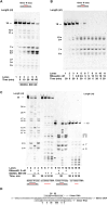
RNaseH2 and DDX3X process R-loops with different digestion patterns. (A) Time course of recombinant human DDX3X incubated in the presence of substrate 1. Lane 1, control reaction in the absence of DDX3X. (B) Increasing concentrations of recombinant human RNaseH2 were incubated for the indicated times in the presence of substrate 1. Lane 1, control reaction in the absence of RNaseH2. (C) Comparison of the nuclease activities of DDX3X (lanes 3, 4, 10, 11) and RNaseH2 (lanes 2, 4, 10, 12) either alone or in combination, on the synthetic R-loop substrates 1 (lanes 10–12) and 2 (lanes 2–4), and of DDX3X alone on the 15-mer (lanes 14–16) and 30-mer (lanes 6–8) ssRNA substrates. Lanes 1, 5, 9, 13, control reactions in the absence of enzymes. In all panels, asterisks indicate the major digested products along with the respective nt lengths. (D) Schematic diagram of the major cut sites, indicated by arrows, of RNaseH2 on the RNA strand of substrates 1 (top) and 2 (bottom).
Figure 2.
Figure 2.
Stimulation of R-loop processing activity of RNaseH2 by DDX3X. (A) Increasing concentrations of human recombinant DDX3X were incubated with substrate 1, either alone (lanes 3–8) or in combination with a fixed dose of human recombinant RNaseH2 (lanes 9–14). Lane 1, control reaction in the absence of enzymes; Lane 2 control reaction with RNaseH2 alone. Asterisks indicate relevant digestion products as discussed in the text. (B) Increasing concentrations of DDX3X were incubated with a fixed amount of RNaseH2 on substrate 1 (lanes 7–12). Lane 1, control reaction in the absence of enzymes; Lanes 2–3 control reactions with RNaseH2 alone; Lanes 4–6 control reactions with DDX3X alone. Asterisks indicate relevant digestion products as discussed in the text. (C) Quantification of the digested substrate by a fixed amount of RNaseH2 alone (white bar), increasing amounts of DDX3X alone (black bars) or their combination (grey bars). Values are the mean of two independent experiments. Error bars indicate ± S.E. (D) Increase in the RNaseH2 digestion activity from panel C was plotted as a function of DDX3X concentrations. Data were fitted to Eq. (1) to derive the apparent dissociation constant for DDX3X-RNaseH2 interaction. Values are the mean of two independent experiments. Error bars indicate ± S.E.
Figure 3.
Figure 3.
DDX3X stimulates RNaseH2 independently from its ribonuclease activity. (A) Increasing concentrations of human recombinant DDX3X were incubated with substrate 3, either alone (lanes 3–8) or in combination with a fixed dose of human recombinant RNaseH2 (lanes 9–14). Lane 1, control reaction in the absence of enzymes; Lane 2 control reaction with RNaseH2 alone. (B) Quantification of the digested substrate by a fixed amount of RNaseH2 alone (white bar), increasing amounts of DDX3X alone (black bars) or their combination (grey bars). Values are the mean of two independent experiments. Error bars indicate ± S.E. (C) Increase in the RNaseH2 digestion activity from panel B was plotted as a function of DDX3X concentrations. Data were fitted to Eq. (1) to derive the apparent dissociation constant for DDX3X-RNaseH2 interaction. Values are the mean of two independent experiments. Error bars indicate ± S.E. (D) RNaseH2 was incubated on substrate 1 either alone (lane 2), or in combination with DDX3Xwt (lane 4) or the DDX3X(132–607) deletion mutant (lanes 7, 8). Lane 1, control reaction in the absence of enzyme; Lane 3 control reaction with DDX3Xwt alone; Lanes 5, 6, control reactions with DDX3X(132–607) alone. (E) As in panel D but in the presence of substrate 3.
Figure 4.
Figure 4.
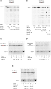
DDX3X helicase activity on R-loops is inhibited by RNaseH2 and is dispensable for its stimulation. (A) Helicase activity of DDX3X on substrate 1 in the absence (lane 2) or in the presence (lanes 3, 4) of ATP. Lane 1, control reaction in the absence of DDX3X; Lane 5, 15-mer ssRNA strand alone as marker for the displaced products. (B) Increasing amounts of DDX3X were titrated in a helicase assay on substrate 1, either alone (lanes 3–5) or in combination with a fixed amount of RNaseH2 (lanes 6–8). Lane 1, control reaction in the absence of enzymes; Lane 2, control reaction with RNaseH2 alone, Lane 9, 15-mer ssRNA strand alone as marker for the displaced products. (C) As in panel B, but in the presence of the DDX3X(132–607) deletion mutant. (D) As in panel B, but in the presence of DDX3X(DADA) mutant. (E) Nuclease activity of DDX3Xwt and mutants either alone (lanes 3, 5, 7) or in combination with RNaseH2 (lanes 4, 6, 8). Lane 1, control reaction in the absence of enzymes; Lane 2, control reaction with RNaseH2 alone.
Figure 5.
Figure 5.
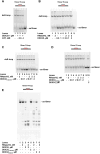
DDX3X physically interacts with the RNaseH2 trimeric complex in an in vitro assay. (A) MBP-tagged DDX3X was incubated with the affinity beads either alone (lanes 2–3) or with human recombinant RNaseH2 (lane 4). Bound proteins were eluted by TEV digestion (lanes 3, 4) and analyzed by Western blot with anti-DDX3X Abs. Lane 1, positive control mixture for purified MBP-DDX3X and DDX3X.; Lane 5, control beads incubated with RNaseH2 alone. (B) Same samples of panel A but analyzed by Western blot with anti-RNaseH2B Abs. Lane 1, positive control for RNaseH2B; Lanes 4, affinity beads incubated with RNaseH2 alone; Lane 5, affinity beads incubated with MBP-DDX3X and RNaseH2 and eluted by TEV digestion. (C) Same samples of panel A and B but analyzed by Western blot with anti-RNaseH2A (lanes 1–5) or anti RNaseH2C (lanes 7–10) Abs. Lanes 1, 7, positive controls for RNaseH2 A and C; Lanes 5, 10, affinity beads incubated with MBP-DDX3X and RNaseH2 and eluted by TEV digestion. (D) MBP-tagged DDX3X was incubated with the affinity beads either alone (lanes 4, 7) or with human recombinant RNaseH2B or RNAseH2C subunits (lanes 8–9). Proteins were eluted by TEV digestion (lanes 7–9) and analyzed by Western blot with anti-DDX3X Abs. Lane 1, positive control mixture for purified MBP-DDX3X and DDX3X, Lane 2, input loaded on MBP affinity beads. Lane 3, flow-through; Lanes 5 and 6, control beads incubated with RNaseH2B or H2C alone. (E) Western blot with anti RNaseH2C (lanes 1–3) or anti RNaseH2B (lanes 5–7) Abs of the corresponding samples shown in panel D. Lanes 1, 5, positive controls for RNaseH2 B and C subunits.
Figure 6.
Figure 6.
DDX3X physically interacts with the RNaseH2 trimeric complex in cell-based assays. (A) Cell lysates of U2OS cells transfected with Halo-DDX3X plasmid (lane 1, 10% of the total) or Halo empty vector (lane 2, 10% of the total). After incubation with Halo-link resin and TEV digestion, fractions were analyzed in Western blot with antibodies specific for DDX3X or RNaseH2 B and C subunits. Lane 3, pull-down after TEV digestion in control lysate transfected with empty vector; Lane 4, pull-down after TEV digestion in lysate transfected with Halo-DDX3X; Lanes 5 and 6, control lysates of U2OS and A549 cells transfected with Halo-DDX3X, respectively; Lane 7, control lysate of untreated U2OS cells; Lane 8, control lysate of U2OS cells silenced with siRNA RNaseH2A and B. (B) Luminescent pull-down assay. Light units obtained are the means ± SEM of three independent experiments. Nluc-N is the positive control, while Nluc-TGM2 and Nluc alone are the negative controls.
Figure 7.
Figure 7.
The B and C subunits of RNaseH2 inhibit DDX3X activities on R-loops. (A) A fixed concentration of human recombinant DDX3X was incubated with substrate 1 either alone (lane 4) or in the presence of increasing concentrations of human recombinant RNaseH2B (lanes 5–7) or H2C (lanes 8–10) subunits. Lane 1, control reaction in the absence of proteins; Lane 2, control reaction in the presence of RNaseH2B; Lane 3, as in lane 2 but in the presence of RNaseH2C. (B) Increasing concentrations of a mixture of RNaseH2B and H2C subunits at equimolar ratios were incubated with substrate 1 either alone (lanes 4–6) or in the presence of a fixed amount of DDX3X (lanes 8–10). Lane 1, control reaction in the absence of proteins; Lane 2, control reaction in the presence of RNaseH2B, Lane 3, as in lane 2 but in the presence of RNaseH2C; Lane 7, reaction with DDX3X alone; Lane 11, control reaction with trimeric RNaseH2; Lanes 12, 13, control reactions with BSA alone or in combination with DDX3X. (C) Helicase activity of DDX3X alone (lane 2) or in combination with increasing amounts of RNaseH2B subunit (lanes 4–8) on substrate 1. Lane 1, control reaction in the absence of proteins, Lane 3, control reaction in the presence of RNaseH2B alone; Lane 9, 15-mer ssRNA oligonucleotide loaded as marker for the displaced products. (D) As in panel C but in the presence of the RNaseH2C subunit. (E). DDX3X alone (lane 2), or increasing concentrations of a mixture of RNaseH2B and H2C subunits at equimolar ratios (lanes 3–5), or a combination of both (lanes 6–8) were incubated on substrate 1 in a helicase assay. Lane 1, control reaction in the absence of proteins; Lane 9, 15-mer ssRNA oligonucleotide loaded as marker for the displaced products.
Figure 8.
Figure 8.
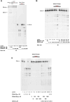
DDX3X stimulation is specific for RNaseH2 catalytic activity. (A) Increasing concentrations of RNaseH1 were titrated in the presence of substrate 3 (lanes 4–8) or substrate 1 (lanes 11–14). Lanes 1, 8: control reactions in the absence of enzymes; Lanes 2, 3, 9, 10: control reactions with two concentrations of RNaseH2. (B) Increasing concentrations of DDX3X were titrated on substrate 1 in the absence (lanes 3–8) or in the presence (lanes 9–14) of a fixed amount of RNaseH1. Lane 1, control reaction in the absence of enzymes; Lane 2, control reaction with RNAseH1 alone. (C) Increasing amounts of RNaseH2(KD) catalytically dead mutant were titrated on substrate 1 in the absence (lanes 6–11) or in the presence (Lanes 12–15) of fixed amounts of DDX3X. Lane 1, control reaction in the absence of enzymes; Lanes 2–5, control reaction with RNaseH2wt alone; Lanes 16–17, control reaction with RNaseH2 in the presence of DDX3X.
Figure 9.
Figure 9.
Silencing of DDX3X in human cells causes accumulation of genomic R-loops and phosphorylated RPA foci. (A) Representative immunofluorescence images obtained using antibodies against RNA:DNA hybrids (S9.6, red) and pRPA (green) after transfection of U2OS cells with control (si-control), DDX3X (siDDX3X), RNaseH2 (siRNaseH2A + siRNAseH2B) or DDX3X and RNaseH2 (siDDX3X + siRNaseH2A + siRNaseH2B) siRNAs. Treatment with RNaseH + RNaseIII + RNaseT1 was used to exclude unspecific S9.6 signals. Scale bars, 10 μm. (B) Quantification of mean nuclear intensity of phospho-RPA and S9.6 foci of control and DDX3X, RNaseH2A + B and DDX3x + RNaseH2A + B-depleted U2OS cells. Median with interquartile range (black lines) are indicated. P values were calculated using two-tailed unpaired Student's t test. *P < 0.05; **P < 0.01; ***P < 0.001; ****P < 0.0001; ns, not significant (P < 0.05). At least 300 nuclei from three different experiments were analyzed. (C) Quantification of RNA:DNA hybrid Dot-blot of genomic DNA ± RNase H for all the siRNA conditions. S9.6 signals are normalized to dsDNA signal. P values were calculated using two-tailed unpaired Student's t test (means ± SEM; n ≥ 2).

References

    1. Petereman E., Lam L., Zou L. Sources, resolution and physiological relevance of R-loops and RNA-DNA hybrids. Nat. Rev. Mol. Cell Biol. 2022; 23:521–540. - PubMed
    1. Brickner J., Garzon J.L., Cimprich K.A. Walking a tightrope: the complex balancing act of R-loops in genome stability. Mol. Cell. 2022; 82:2267–2297. - PMC - PubMed
    1. Lee D.Y., Clayton D.A.. Initiation of mitochondrial DNA replication by transcription and R-loop processing. J. Biol. Chem. 1998; 273:30614–30621. - PubMed
    1. Crossley M.P., Bocek M., Cimprich K.A.. R-loops as cellular regulators and genomic threats. Mol. Cell. 2019; 73:398–411. - PMC - PubMed
    1. Castillo-Guzman D., Chédin F.. Defining R-loop classes and their contributions to genome instability. DNA Repair (Amst.). 2021; 106:103182. - PMC - PubMed

LinkOut - more resources