Review
. 2024 Aug;312(2):e240122.
doi: 10.1148/radiol.240122.

A Lexicon for First-Trimester US: Society of Radiologists in Ultrasound Consensus Conference Recommendations

Affiliations
Review

A Lexicon for First-Trimester US: Society of Radiologists in Ultrasound Consensus Conference Recommendations

Shuchi K Rodgers et al. Radiology. 2024 Aug.

Abstract

The Society of Radiologists in Ultrasound convened a multisociety panel to develop a first-trimester US lexicon based on scientific evidence, societal guidelines, and expert consensus that would be appropriate for imagers, clinicians, and patients. Through a modified Delphi process with consensus of at least 80%, agreement was reached for preferred terms, synonyms, and terms to avoid. An intrauterine pregnancy (IUP) is defined as a pregnancy implanted in a normal location within the uterus. In contrast, an ectopic pregnancy (EP) is any pregnancy implanted in an abnormal location, whether extrauterine or intrauterine, thus categorizing cesarean scar implantations as EPs. The term pregnancy of unknown location is used in the setting of a pregnant patient without evidence of a definite or probable IUP or EP at transvaginal US. Since cardiac development is a gradual process and cardiac chambers are not fully formed in the first trimester, the term cardiac activity is recommended in lieu of 'heart motion' or 'heartbeat.' The terms 'living' and 'viable' should also be avoided in the first trimester. 'Pregnancy failure' is replaced by early pregnancy loss (EPL). When paired with various modifiers, EPL is used to describe a pregnancy in the first trimester that may or will not progress, is in the process of expulsion, or has either incompletely or completely passed. © RSNA and Elsevier, 2024 Supplemental material is available for this article. This article is a simultaneous joint publication in Radiology and American Journal of Obstetrics & Gynecology. All rights reserved. The articles are identical except for minor stylistic and spelling differences in keeping with each journal's style. Either version may be used in citing this article. See also the editorial by Scoutt and Norton in this issue.


Conflict of interest statement

Disclosures of conflicts of interest: S.K.R. Book royalties from Elsevier. M.M.H. Payment for lectures, travel, or visiting professorship from Massachusetts General Hospital/Brigham Women’s Hospital, University of Toronto, McMaster University, World Class CME, and Hoag Medical Group; section editor for RadioGraphics. P.M.D. No relevant relationships. M.C.F. Payment for panel discussion from GE HealthCare. A. Kennedy Royalties and writer’s fees from Elsevier; honoraria from World Class CME and University of California Davis; support for travel from the RSNA; secretary of the Society of Radiologists in Ultrasound. R.A. No relevant relationships. K.B. Patents planned, issued, or pending with Kristyn Brandi MD MPH LLC. L.D. No relevant relationships. S.K.H. Grants from the National Center for Advancing Translational Sciences at the National Institutes of Health; payment for presentations from Organon; payment for expert testimony from Huff Powell Bailey; support for attending meeting from the American College of Obstetricians and Gynecologists (ACOG); section secretary of the Pennsylvania ACOG. A. Kamaya Book royalties from Elsevier. A. Koyama No relevant relationships. P.C.L. Honoraria for editor role from RoshReview (now Blueprint); support to attend meeting from Individuals with Intellectual or Development Disability Engaged, Aligned and Leading project workgroup funded by the Patient-Centered Outcomes Research Institute; secretary-treasurer of the New York Chapter of the American College of Emergency Physicians and past chair of the American College of Emergency Physicians Emergency Ultrasound Section. K.E.M. No relevant relationships. T.M. Book royalties from Elsevier. S.G.O. No relevant relationships. K.O. No relevant relationships. R.S. Honoraria and support for travel to meetings from World Class CME. S.S. Grants from the National Institutes of Health, AbbVie, and Organon. L.M.S. No relevant relationships.

Figures

None
Graphical abstract
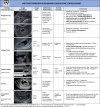
General terms. Lexicon terms (bolded and/or italicized) applicable to
pregnancy but not specific to imaging are listed in this table.
Figure 1:
General terms. Lexicon terms (bolded and/or italicized) applicable to pregnancy but not specific to imaging are listed in this table.
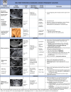
Early development. Lexicon terms (bolded and/or italicized) in this
table relate to structures visualized at US in early pregnancy development.
Terms to avoid are in single quotation marks. Terms specific to
multigestation pregnancies, such as chorionicity and amnionicity, are beyond
the scope of this lexicon.
Figure 2:
Early development. Lexicon terms (bolded and/or italicized) in this table relate to structures visualized at US in early pregnancy development. Terms to avoid are in single quotation marks. Terms specific to multigestation pregnancies, such as chorionicity and amnionicity, are beyond the scope of this lexicon.
Intradecidual sign. Transvaginal sagittal grayscale US image in a
34-year-old pregnant patient shows a 4-mm empty gestational sac (GS) (solid
arrow) in the anterior endometrium. The location of the GS to one side of
the central hyperechoic line (dotted arrows) representing the opposed
innermost layers of decidualized endometrium confirms it is within
endometrium and not the endometrial cavity, hence the name intradecidual
sign. This sign is helpful when seen to diagnose a probable intrauterine
pregnancy (IUP) before visualization of the yolk sac. Three days later, the
yolk sac was visualized (not shown), allowing for the diagnosis of a
definite IUP.
Figure 3:
Intradecidual sign. Transvaginal sagittal grayscale US image in a 34-year-old pregnant patient shows a 4-mm empty gestational sac (GS) (solid arrow) in the anterior endometrium. The location of the GS to one side of the central hyperechoic line (dotted arrows) representing the opposed innermost layers of decidualized endometrium confirms it is within endometrium and not the endometrial cavity, hence the name intradecidual sign. This sign is helpful when seen to diagnose a probable intrauterine pregnancy (IUP) before visualization of the yolk sac. Three days later, the yolk sac was visualized (not shown), allowing for the diagnosis of a definite IUP.
Double decidual sac sign. Transvaginal transverse grayscale US image
in a 27-year-old pregnant patient at 5 weeks 3 days shows two concentric
echogenic rings around an oval fluid collection representing the double
decidual sac sign. The inner echogenic ring (arrows) corresponds to
trophoblastic chorion and decidua capsularis. The outer echogenic ring
(arrowheads) represents decidua vera and endometrial lining. The double
decidual sac sign is more specific for an intrauterine pregnancy than the
intradecidual sign when the gestational sac is empty.
Figure 4:
Double decidual sac sign. Transvaginal transverse grayscale US image in a 27-year-old pregnant patient at 5 weeks 3 days shows two concentric echogenic rings around an oval fluid collection representing the double decidual sac sign. The inner echogenic ring (arrows) corresponds to trophoblastic chorion and decidua capsularis. The outer echogenic ring (arrowheads) represents decidua vera and endometrial lining. The double decidual sac sign is more specific for an intrauterine pregnancy than the intradecidual sign when the gestational sac is empty.
Pregnancy location. The location of a pregnancy is divided into
normal, abnormal, and unknown. Lexicon terms are bolded and/or italicized,
and terms to avoid are in single quotation marks. The essential word in the
definition of intrauterine pregnancy and ectopic pregnancy (EP) is
implanted, which helps differentiate pregnancies that are temporarily
located in the lower uterine segment. This definition also further clarifies
abnormal intrauterine implantation sites as EPs.
Figure 5:
Pregnancy location. The location of a pregnancy is divided into normal, abnormal, and unknown. Lexicon terms are bolded and/or italicized, and terms to avoid are in single quotation marks. The essential word in the definition of intrauterine pregnancy and ectopic pregnancy (EP) is implanted, which helps differentiate pregnancies that are temporarily located in the lower uterine segment. This definition also further clarifies abnormal intrauterine implantation sites as EPs.
Normal intrauterine pregnancy (IUP) variant: eccentrically located
gestational sac (GS) completely surrounded by endometrium. (A) Transvaginal
transverse grayscale and (B) coronal reformatted three-dimensional US image
in a 36-year-old pregnant patient shows an off-midline (leftward) GS (arrow)
at 5 weeks 4 days with yolk sac and embryo. The GS is completely surrounded
by endometrium and may be reported as an IUP without further description. If
desired, the user may describe as an eccentrically located GS completely
surrounded by endometrium but should conclude as an IUP to obviate concern
for an interstitial ectopic pregnancy. The terms ‘angular’ or
‘cornual pregnancy’ should be avoided. Coronal reformatted
three-dimensional US may help confirm a GS is located within endometrium, as
demonstrated in this case.
Figure 6:
Normal intrauterine pregnancy (IUP) variant: eccentrically located gestational sac (GS) completely surrounded by endometrium. (A) Transvaginal transverse grayscale and (B) coronal reformatted three-dimensional US image in a 36-year-old pregnant patient shows an off-midline (leftward) GS (arrow) at 5 weeks 4 days with yolk sac and embryo. The GS is completely surrounded by endometrium and may be reported as an IUP without further description. If desired, the user may describe as an eccentrically located GS completely surrounded by endometrium but should conclude as an IUP to obviate concern for an interstitial ectopic pregnancy. The terms ‘angular’ or ‘cornual pregnancy’ should be avoided. Coronal reformatted three-dimensional US may help confirm a GS is located within endometrium, as demonstrated in this case.
Specific normal and abnormal pregnancy location sites. Schematic
illustration of normal pregnancy implantation sites on the left half of the
uterine diagram and abnormal implantation sites on the right. Representative
round icons indicate the implantation site with corresponding letters to
lexicon terms in the box. Of note, it is optional to further describe a
tubal ectopic pregnancy location as isthmic, infundibular, or ampullary when
the precise location is clear at US.
Figure 7:
Specific normal and abnormal pregnancy location sites. Schematic illustration of normal pregnancy implantation sites on the left half of the uterine diagram and abnormal implantation sites on the right. Representative round icons indicate the implantation site with corresponding letters to lexicon terms in the box. Of note, it is optional to further describe a tubal ectopic pregnancy location as isthmic, infundibular, or ampullary when the precise location is clear at US.
Heterotopic pregnancy. Transvaginal transverse grayscale US image in a
27-year-old pregnant patient shows an early intrauterine pregnancy (IUP)
(solid arrow) containing a yolk sac and a tubal ring of ectopic pregnancy
(EP) (arrowhead) also containing a yolk sac. The coexistence of an IUP and
EP is termed heterotopic pregnancy. The left ovary contains a corpus luteum
(dotted arrow) and is seen in the center. The rim of chorionic tissue in the
IUP and EP is hyperechoic, whereas in contrast, the corpus luteum is
hypoechoic. Echogenicity can help distinguish a tubal ring from a corpus
luteum in some cases. UT = uterus.
Figure 8:
Heterotopic pregnancy. Transvaginal transverse grayscale US image in a 27-year-old pregnant patient shows an early intrauterine pregnancy (IUP) (solid arrow) containing a yolk sac and a tubal ring of ectopic pregnancy (EP) (arrowhead) also containing a yolk sac. The coexistence of an IUP and EP is termed heterotopic pregnancy. The left ovary contains a corpus luteum (dotted arrow) and is seen in the center. The rim of chorionic tissue in the IUP and EP is hyperechoic, whereas in contrast, the corpus luteum is hypoechoic. Echogenicity can help distinguish a tubal ring from a corpus luteum in some cases. UT = uterus.
Corpus luteum. (A) Transvaginal sagittal grayscale and (B) color
Doppler US image in a 31-year-old pregnant patient shows a round,
hypoechoic, thick-walled structure (dotted arrow) with a central cystic
space (*) and peripheral vascularity (arrowheads), characteristic of
a corpus luteum. This corpus luteum arises in an exophytic fashion from the
right ovary (OV), which can mimic a tubal ring of ectopic pregnancy. A
helpful feature to diagnose a corpus luteum is a claw sign (solid arrows) of
partially surrounding ovarian parenchyma, which confirms an ovarian
origin.
Figure 9:
Corpus luteum. (A) Transvaginal sagittal grayscale and (B) color Doppler US image in a 31-year-old pregnant patient shows a round, hypoechoic, thick-walled structure (dotted arrow) with a central cystic space (*) and peripheral vascularity (arrowheads), characteristic of a corpus luteum. This corpus luteum arises in an exophytic fashion from the right ovary (OV), which can mimic a tubal ring of ectopic pregnancy. A helpful feature to diagnose a corpus luteum is a claw sign (solid arrows) of partially surrounding ovarian parenchyma, which confirms an ovarian origin.
Interstitial ectopic pregnancy (EP). (A) Transvaginal transverse
grayscale and (B) coronal reformatted three-dimensional US image in a
29-year-old pregnant patient shows a gestational sac (GS) (calipers) at 5
weeks 3 days. There is intervening myometrium (solid arrow) between the GS
and endometrium (*). A claw sign (dotted arrows) of myometrial tissue
confirms the pregnancy is implanted in the interstitial (intramyometrial)
segment of the tube, termed an interstitial EP. The term ‘cornual
EP’ should be avoided. Coronal reformatted three-dimensional US may
better demonstrate an interstitial EP separate from the endometrium, as in
this case. Diam = diameter.
Figure 10:
Interstitial ectopic pregnancy (EP). (A) Transvaginal transverse grayscale and (B) coronal reformatted three-dimensional US image in a 29-year-old pregnant patient shows a gestational sac (GS) (calipers) at 5 weeks 3 days. There is intervening myometrium (solid arrow) between the GS and endometrium (*). A claw sign (dotted arrows) of myometrial tissue confirms the pregnancy is implanted in the interstitial (intramyometrial) segment of the tube, termed an interstitial EP. The term ‘cornual EP’ should be avoided. Coronal reformatted three-dimensional US may better demonstrate an interstitial EP separate from the endometrium, as in this case. Diam = diameter.
Interstitial line sign. Transvaginal transverse grayscale US image in
a 33-year-old pregnant patient shows a right interstitial ectopic pregnancy
(EP) (solid arrow) at 6 weeks 0 days. In addition to the characteristic
sonographic findings of an interstitial EP shown in Figure 10, there is a
thin echogenic line (dotted arrows) representing the interstitial segment of
the tube. This line connects the endometrium to the ectopic gestational sac
and is called the interstitial line sign.
Figure 11:
Interstitial line sign. Transvaginal transverse grayscale US image in a 33-year-old pregnant patient shows a right interstitial ectopic pregnancy (EP) (solid arrow) at 6 weeks 0 days. In addition to the characteristic sonographic findings of an interstitial EP shown in Figure 10, there is a thin echogenic line (dotted arrows) representing the interstitial segment of the tube. This line connects the endometrium to the ectopic gestational sac and is called the interstitial line sign.
Cervical ectopic pregnancy (EP). (A) Transvaginal sagittal grayscale
US of the uterus and (B) high-resolution US image of the cervix in a
31-year-old pregnant patient at 6 weeks 3 days with vaginal bleeding. A
gestational sac (GS) (solid arrow) is seen containing an embryo with cardiac
activity (not shown) implanted in the cervix. Mixed-echogenicity material
(arrowhead) representing blood products expands the endometrial cavity. The
location of the GS eccentric to the endocervical canal (dotted arrows) and
cardiac activity help make the diagnosis of a cervical EP. The term
‘cervical pregnancy’ should be avoided.
Figure 12:
Cervical ectopic pregnancy (EP). (A) Transvaginal sagittal grayscale US of the uterus and (B) high-resolution US image of the cervix in a 31-year-old pregnant patient at 6 weeks 3 days with vaginal bleeding. A gestational sac (GS) (solid arrow) is seen containing an embryo with cardiac activity (not shown) implanted in the cervix. Mixed-echogenicity material (arrowhead) representing blood products expands the endometrial cavity. The location of the GS eccentric to the endocervical canal (dotted arrows) and cardiac activity help make the diagnosis of a cervical EP. The term ‘cervical pregnancy’ should be avoided.
Ovarian ectopic pregnancy (EP). Transvaginal sagittal grayscale US
image in a 35-year-old pregnant patient at 6 weeks 4 days shows a
gestational sac (GS) (arrow) containing a yolk sac and embryo with cardiac
activity (not shown) within the ovary (Ov) (calipers), diagnostic of an
ovarian EP. A peripheral follicle is present in the ovary (arrowhead). To
avoid misdiagnosing a corpus luteum for the rare ovarian EP, a yolk sac or
embryo should be present in the intraovarian thick-walled cystic structure
representing the GS. UT = uterus.
Figure 13:
Ovarian ectopic pregnancy (EP). Transvaginal sagittal grayscale US image in a 35-year-old pregnant patient at 6 weeks 4 days shows a gestational sac (GS) (arrow) containing a yolk sac and embryo with cardiac activity (not shown) within the ovary (Ov) (calipers), diagnostic of an ovarian EP. A peripheral follicle is present in the ovary (arrowhead). To avoid misdiagnosing a corpus luteum for the rare ovarian EP, a yolk sac or embryo should be present in the intraovarian thick-walled cystic structure representing the GS. UT = uterus.
Pregnancy of unknown location (PUL). The original definition of PUL is
maintained; however, it is clarified in the lexicon to clearly state that
there should be no evidence of probable or definite intrauterine pregnancy
(IUP) or ectopic pregnancy (EP) to qualify as a PUL. An empty gestational
sac is considered a probable pregnancy whether implanted in a normal
location (IUP) or abnormal location (EP) and should not be termed a
PUL.
Figure 14:
Pregnancy of unknown location (PUL). The original definition of PUL is maintained; however, it is clarified in the lexicon to clearly state that there should be no evidence of probable or definite intrauterine pregnancy (IUP) or ectopic pregnancy (EP) to qualify as a PUL. An empty gestational sac is considered a probable pregnancy whether implanted in a normal location (IUP) or abnormal location (EP) and should not be termed a PUL.
Early pregnancy loss (EPL). There are five main categories of EPL:
concerning for, diagnostic of, in progress, incomplete, and completed.
Enhanced myometrial vascularity (EMV) is included in the lexicon since
increased myometrial vascularity deep to a prior implantation site is
commonly confused with other rare entities, such as an arteriovenous fistula
and arteriovenous malformation, which may lead to unnecessary
work-up.
Figure 15:
Early pregnancy loss (EPL). There are five main categories of EPL: concerning for, diagnostic of, in progress, incomplete, and completed. Enhanced myometrial vascularity (EMV) is included in the lexicon since increased myometrial vascularity deep to a prior implantation site is commonly confused with other rare entities, such as an arteriovenous fistula and arteriovenous malformation, which may lead to unnecessary work-up.
Poor prognosticators: calcified yolk sac and expanded amnion sign.
Transvaginal sagittal grayscale US image in a 27-year-old pregnant patient
shows an 8-mm embryo (calipers) without cardiac activity (M-mode not shown)
sufficient for the interpretation of diagnostic of EPL. Additional poor
prognosticators include a calcified yolk sac (arrow) and an enlarged
amniotic cavity (arrowhead) relative to the crown-rump length (CRL) of the
embryo, called the expanded amnion sign. These additional observations, on
their own, are only concerning for EPL. GA = gestational age.
Figure 16:
Poor prognosticators: calcified yolk sac and expanded amnion sign. Transvaginal sagittal grayscale US image in a 27-year-old pregnant patient shows an 8-mm embryo (calipers) without cardiac activity (M-mode not shown) sufficient for the interpretation of diagnostic of EPL. Additional poor prognosticators include a calcified yolk sac (arrow) and an enlarged amniotic cavity (arrowhead) relative to the crown-rump length (CRL) of the embryo, called the expanded amnion sign. These additional observations, on their own, are only concerning for EPL. GA = gestational age.
Summary of major lexicon changes highlighting terms to use. The major
changes from currently used terminology to describe sonographic findings in the
first trimester are (a) early pregnancy loss in lieu of ‘failure’;
(b) cardiac activity in lieu of ‘heart motion’; and (c) defining
ectopic pregnancy as an abnormal implantation site. The terms
‘live,’ ‘living,’ and ‘viable’ are
commonly used terms to describe cardiac activity. However, as these terms may be
misleading, they are best avoided in the first trimester.
Figure 17:
Summary of major lexicon changes highlighting terms to use. The major changes from currently used terminology to describe sonographic findings in the first trimester are (a) early pregnancy loss in lieu of ‘failure’; (b) cardiac activity in lieu of ‘heart motion’; and (c) defining ectopic pregnancy as an abnormal implantation site. The terms ‘live,’ ‘living,’ and ‘viable’ are commonly used terms to describe cardiac activity. However, as these terms may be misleading, they are best avoided in the first trimester.
Summary of major lexicon changes highlights terms to avoid. Equally
important as terms to use are those terms that are best avoided since they are
obsolete or confusing (single quotation marks). This is accompanied by
recommended lexicon terms (bold and italicized) to use instead.
Figure 18:
Summary of major lexicon changes highlights terms to avoid. Equally important as terms to use are those terms that are best avoided since they are obsolete or confusing (single quotation marks). This is accompanied by recommended lexicon terms (bold and italicized) to use instead.

References

    1. AIUM practice parameter for the performance of standard diagnostic obstetric ultrasound . J Ultrasound Med 2024. ; 43 ( 6 ): E20 – E32 . - PubMed
    1. Barnhart K , van Mello NM , Bourne T , et al . Pregnancy of unknown location: a consensus statement of nomenclature, definitions, and outcome . Fertil Steril 2011. ; 95 ( 3 ): 857 – 866 . - PMC - PubMed
    1. Doubilet PM, Benson CB, Bourne T, et al. ; Society of Radiologists in Ultrasound Multispecialty Panel on Early First Trimester Diagnosis of Miscarriage and Exclusion of a Viable Intrauterine Pregnancy . Diagnostic criteria for nonviable pregnancy early in the first trimester. N Engl J Med 2013;369(15):1443–1451. - PubMed
    1. Kolte AM , Bernardi LA , Christiansen OB , et al. ; ESHRE Special Interest Group, Early Pregnancy . Terminology for pregnancy loss prior to viability: a consensus statement from the ESHRE early pregnancy special interest group . Hum Reprod 2015. ; 30 ( 3 ): 495 – 498 . - PubMed
    1. Kirk E , Ankum P , Jakab A , et al. ; ESHRE Working Group on Ectopic Pregnancy . Terminology for describing normally sited and ectopic pregnancies on ultrasound: ESHRE recommendations for good practice . Hum Reprod Open 2020. ; 2020 ( 4 ): a055 . - PMC - PubMed

LinkOut - more resources