

Changes in MRI head motion across development: typical development and ADHD
- PMID: 39190098
- PMCID: PMC11582210
- DOI: 10.1007/s11682-024-00910-w
Changes in MRI head motion across development: typical development and ADHD
Abstract
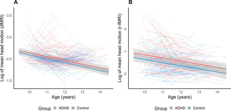
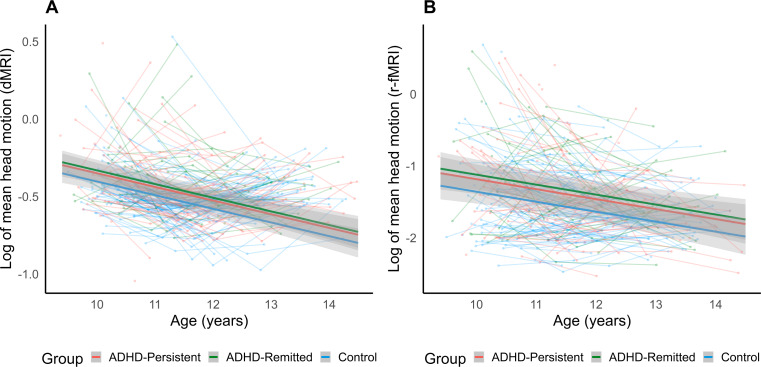
Head motion is a major confounding variable for magnetic resonance imaging (MRI) analysis, and is commonly seen in individuals with neurodevelopmental disorders such as attention deficit hyperactivity disorder (ADHD). This study investigated the trajectory of change in head motion in typically developing children and children with ADHD, and examined possible altered trajectories in head motion between children with remitted and persistent ADHD. 105 children with ADHD and 84 controls completed diffusion and resting-state functional MRI scans at up to three waves over ages 9-14 years. In-scanner head motion was calculated using framewise displacement, and longitudinal trajectories analyzed using generalized additive mixed modelling. Results revealed a significant age effect on framewise displacement where head motion decreased as age increased during both diffusion (p < .001) and resting-state functional MRI (p < .001). A significant effect of group was also observed; children with ADHD displayed greater framewise displacement than controls over the age range (diffusion MRI p = .036, functional MRI p = .004). Further analyses revealed continued elevation in head motion in children in remission from ADHD (diffusion MRI p = .020, functional MRI p = .011) compared to controls. Rates of change in head motion did not significantly differ between diagnostic groups. Findings indicate a critical link between in-scanner head motion and developmental age within children regardless of ADHD diagnosis, important to consider in studies of neurodevelopment. Findings also suggest change in head motion with age does not differ between individuals with remitted and persistent ADHD, adding further evidence that behavioral manifestations of ADHD may continue despite clinical remission.
Keywords: ADHD; Diffusion MRI; Longitudinal development; Motion artifact; Resting-state functional MRI.
© 2024. The Author(s).
Conflict of interest statement
Declarations. Competing interests: The authors declare no competing interests. Ethics approval: The Neuroimaging of the Children’s Attention project was approved by the Royal Children’s Hospital Human Research Ethics Committee in Melbourne. Consent to participate: All participating families provided written informed consent prior to data collection. Competing interests: None of the authors have a conflict of interest to declare.
Figures
Similar articles
-
Developmental brain trajectories in children with ADHD and controls: a longitudinal neuroimaging study.BMC Psychiatry. 2016 Mar 11;16:59. doi: 10.1186/s12888-016-0770-4. BMC Psychiatry. 2016. PMID: 26969310 Free PMC article.
-
Head Motion During MRI Predicted by out-of-Scanner Sustained Attention Performance in Attention-Deficit/Hyperactivity Disorder.J Atten Disord. 2021 Aug;25(10):1429-1440. doi: 10.1177/1087054720911988. Epub 2020 Mar 19. J Atten Disord. 2021. PMID: 32189534
-
Functional and structural brain network development in children with attention deficit hyperactivity disorder.Hum Brain Mapp. 2023 Jun 1;44(8):3394-3409. doi: 10.1002/hbm.26288. Epub 2023 Mar 29. Hum Brain Mapp. 2023. PMID: 36988503 Free PMC article.
-
Research Review: Diffusion tensor imaging studies of attention-deficit/hyperactivity disorder: meta-analyses and reflections on head motion.J Child Psychol Psychiatry. 2018 Mar;59(3):193-202. doi: 10.1111/jcpp.12778. Epub 2017 Jul 3. J Child Psychol Psychiatry. 2018. PMID: 28671333
-
Revealing the Structural Neural Circuitry of Attention Deficit Hyperactivity Disorder With Diffusion MRI: Implications for Future Diagnosis and Treatment.AJR Am J Roentgenol. 2018 Apr;210(4):731-733. doi: 10.2214/AJR.17.18983. Epub 2018 Jan 11. AJR Am J Roentgenol. 2018. PMID: 29323553 Review.
Cited by
-
Age- and sex-related differences in social competence and emotion labeling in pre-adolescence.Dev Cogn Neurosci. 2025 Jan;71:101503. doi: 10.1016/j.dcn.2024.101503. Epub 2024 Dec 24. Dev Cogn Neurosci. 2025. PMID: 39733501 Free PMC article.
References
-
- Anderson, A. W., & Gore, J. C. (1994). Analysis and correction of motion artifacts in diffusion weighted imaging. Magnetic Resonance in Medicine, 32(3), 379–387. 10.1002/mrm.1910320313 - PubMed
-
- Aoki, Y., Cortese, S., & Castellanos, F. X. (2018). Research review: Diffusion tensor imaging studies of attention-deficit/hyperactivity disorder: Meta‐analyses and reflections on head motion. Journal of Child Psychology and Psychiatry, 59(3), 193–202. 10.1111/jcpp.12778 - PubMed
-
- Baum, G. L., Roalf, D. R., Cook, P. A., Ciric, R., Rosen, A. F., Xia, C., Elliott, M. A., Ruparel, K., Verma, R., Tunç, B., Gur, R. C., Gur, R. E., Bassett, D. S., & Satterthwaite, T. D. (2018). The impact of in-scanner head motion on structural connectivity derived from diffusion MRI. Neuroimage, 173, 275–286. 10.1016/j.neuroimage.2018.02.041 - PMC - PubMed
-
- Biederman, J., Mick, E., & Faraone, S. V. (2000). Age-dependent decline of symptoms of attention deficit hyperactivity disorder: Impact of remission definition and symptom type. The American Journal of Psychiatry, 157(5), 816–818. 10.1176/appi.ajp.157.5.816 - PubMed
MeSH terms
LinkOut - more resources
Full Text Sources
Medical
