. 2024 Sep 3;121(36):e2409493121.
doi: 10.1073/pnas.2409493121. Epub 2024 Aug 27.

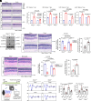
Microglial cGAS-STING signaling underlies glaucoma pathogenesis

Affiliations

Microglial cGAS-STING signaling underlies glaucoma pathogenesis

Yutong Liu et al. Proc Natl Acad Sci U S A. .

Abstract

Characterized by progressive degeneration of retinal ganglion cells (RGCs) and vision loss, glaucoma is the primary cause of irreversible blindness, incurable and affecting over 78 million patients. However, pathogenic mechanisms leading to glaucoma-induced RGC loss are incompletely understood. Unexpectedly, we found that cGAS-STING (2'3'-cyclic GMP-AMP-stimulator of interferon genes) signaling, which surveils displaced double-stranded DNA (dsDNA) in the cytosol and initiates innate immune responses, was robustly activated during glaucoma in retinal microglia in distinct murine models. Global or microglial deletion of STING markedly relieved glaucoma symptoms and protected RGC degeneration and vision loss, while mice bearing genetic cGAS-STING supersensitivity aggravated retinal neuroinflammation and RGC loss. Mechanistically, dsDNA from tissue injury activated microglial cGAS-STING signaling, causing deleterious macroglia reactivity in retinas by cytokine-mediated microglia-macroglia interactions, progressively driving apoptotic death of RGCs. Remarkably, preclinical investigations of targeting cGAS-STING signaling by intraocular injection of TBK1i or anti-IFNAR1 antibody prevented glaucoma-induced losses of RGCs and vision. Therefore, we unravel an essential role of cGAS-STING signaling underlying glaucoma pathogenesis and suggest promising therapeutic strategies for treating this devastating disease.

Keywords: cGAS–STING; glaucoma; microglia; neuroinflammation; vision loss.


Conflict of interest statement

Competing interests statement:The authors declare no competing interest.

Figures

Fig. 1.
Fig. 1.
cGAS–STING signaling is robustly activated in microglia upon ONC. (A) The schematic indicates crush experiments of the optic nerve used in this study. Unless specified in the figures, the samples were harvested at 3 d post crush (dpc) for immunoblotting and 7 dpc for immunofluorescence and qRT-PCR experiments. (B) Immunoblottings detected an increased expression of Iba1 in the injured retinas. (C) Immunofluorescence results showed that both Iba1 and P2ry12 labeled microglia in the retinas and visualized an accumulation of Iba1 (red) and a decrease in P2ry12 (green) in retinas upon ONC. (D) Immunofluorescence visualized aggregated STING proteins (green) in Iba1-positive cells. No STING staining was visible in Sting1−/− mouse retinas. (E) Representative photographs showed a significant accumulation of dsDNA in the injured retinas, revealed by immunofluorescence using an anti-dsDNA antibody. (F) Representative photographs showed mitochondrial-derived dsDNA, revealed by immunofluorescence using the anti-dsDNA (red) and the anti-Tom20 (green) antibody. (G) Immunoblottings detected the robust phosphorylation of TBK1 (S172 residue) and STAT1 (S727 residue) and increased expression of cGAS and STING in retinas at 3 dpc. (H) qRT-PCR detected increased expression of ISGs in the injured retinas at 7 dpc, including Ifit1, Ifit2, Isg15, and Ccl5. Figs. 1–7 and SI Appendix, Figs. S1–S8: n = 3 independent biological repeats unless specified. Data show mean ± SEM, and statistical analyses by ANOVA with Bonferroni correction. [Scale bar, C, 100 μm (50 μm in amplified panel); DF, 10 μm.]
Fig. 2.
Fig. 2.
Genetic ablation of Sting1 alleviates glaucoma-induced loss of RGCs. (A) Representative photographs of H&E staining showed progressive losses of RGCs at 0, 7, 14, and 21 dpc, respectively, revealing that survival ratios of RGCs decreased over time upon ONC. (B and C) STAT1 phosphorylation level (B) and ISG mRNA levels (C) were not responsive in Sting1−/− retinas upon ONC, contradicting those in WT mice, as revealed by immunoblotting and qRT-PCR, respectively. (D–F) Representative photographs of H&E staining (D), their quantitative analyses (E), and the survival ratio of RGCs (F) exhibited a loss of RGCs upon ONC at 7 dpc, which was compromised in Sting1−/− retinas from merely 60.1 to 82.6%. (G–I) Representative photographs (G) and quantitative analyses (H and I) showed fewer apoptotic cells at 28 dpi in Sting1−/− retinas injected with microbeads than in WT mice. (J) Schematic drawing to show F-VEP assays. (K) Representative images of the F-VEP wave of WT and Sting1−/− mice at 7 dpc upon ONC were shown, alongside quantitative analyses of the amplitude and the latency of the N1P1 wave in the F-VEP (Right panel, K). Each dot represents one experiment in C, and each dot represents one mouse in E, F, H, I, and K. (Scale bars in A, D, and G, 100 μm.) ONL, outer nuclear layer; OPL, outer plexiform layer; INL, inner nuclear layer; IPL, inner plexiform layer; GCL, ganglion cells layer.
Fig. 3.
Fig. 3.
Ablation of Sting1 in microglia alleviates glaucoma-induced loss of RGCs. (A) Representative immunoblottings showed that attenuated TBK1 and STAT1 phosphorylation levels were seen in retinas from Sting1fl/fl;Cx3cr1CreERT/+ mice upon ONC. (B–D) Representative photographs of H&E staining (B) and their analyses of RGC numbers (C) and survival ratio (D) showed that cell-specific Sting1 ablation promoted the survival of RGCs upon ONC. (E–G) Representative images of the F-VEP wave of vehicle/tamoxifen-treated Sting1fl/fl;Cx3cr1CreERT/+ mice at 7 dpc upon ONC were shown (E), alongside quantitative analyses of the amplitude (F) and the latency (G) of the N1P1 wave in the F-VEP. Each dot represents one mouse in C, D, F, and G. (Scale bars, B, 100 μm.)
Fig. 4.
Fig. 4.
Supersensitive cGAS–STING signaling (Tbk1S511E/S511E knock-in) aggravates RGC loss in glaucoma mice. (A and B) Immunoblottings (A) and qRT-PCR (B) detected the increased phosphorylation levels of TBK1 and STAT1 and ISG expression in Tbk1S511E/S511E knock-in retinas upon ONC at 3 dpc. (C–E) Representative photographs of H&E staining (C) and their analyses of RGC number (D) and survival ratio (E) showed that the survival of RGCs upon ONC at 7 dpc was markedly worse in Tbk1S511E/S511E knock-in retinas. Each dot represents one experiment in B, and each dot represents one mouse in D and E. (Scale bars, C, 100 μm.)
Fig. 5.
Fig. 5.
cGAS–STING signaling induces Müller glia reactivity and neuroinflammation in ONC-injured retinas. (A) Immunofluorescence imaging showed that ONC-induced accumulation of microglia in vehicle/tamoxifen-treated Sting1fl/fl;Cx3cr1CreERT/+ mouse retinas at 7 dpc upon ONC. (B) Quantitative analyses of the size of each microglia (Iba1-positive cell) in flat-mounted retinas between vehicle and tamoxifen-treated Sting1fl/fl;Cx3cr1CreERT/+ mouse in response to ONC surgery. (C) Upon ONC, Cd68 mRNA levels were compromised in tamoxifen-treated Sting1fl/fl;Cx3cr1CreERT/+ mouse retinas, as revealed by qRT-PCR assays. (D) Upon ONC, Gfap mRNA levels were compromised in tamoxifen-treated Sting1fl/fl;Cx3cr1CreERT/+ mouse retinas, as revealed by qRT-PCR assays. (E) Representative photographs of immunofluorescence against GFAP showed that genetic ablation of Sting1 in microglia markedly inhibited the activation of macroglia (majorly Müller glia) by ONC surgery. (F) qRT-PCR assays showed that Sting1 ablation in microglia robustly inhibited the mRNA expression of proinflammatory cytokines induced by ONC. Each dot represents one experiment in C, D, and F. (Scale bars, A and E, 100 μm.)
Fig. 6.
Fig. 6.
Targeting TBK1 in retinas relieves glaucoma-induced neuroinflammation and RGC loss. (A and B) Immunoblottings and qRT-PCR detected a marked inhibition of TBK1 and STAT1 phosphorylation (A) and ISG induction (B) in retinas by TBK1i, otherwise activated by ONC surgery. (C and D) Representative photographs of H&E staining (C) and their survival ratio (D) showed TBK1i administration effectively promoted RGC survival in ONC-induced retinas, from merely 59.6% to 79.2% at 7 dpc. (E–G) Representative images of the F-VEP wave of vehicles or TBK1i treated mice at 7 dpc upon ONC (E), alongside quantitative analyses of the amplitude (F) and the latency (G) of N1P1 wave in the F-VEP. Each dot represents one experiment in B, and each dot represents one mouse in D, F, and G. (Scale bars, C, 100 μm.)
Fig. 7.
Fig. 7.
Antibody-based neutralization of IFN-I signaling protects RGCs and vision in glaucoma mice. (A and B) Immunoblottings and qRT-PCR detected a noticeable inhibition of STAT1 phosphorylation (A) and ISG induction (B) in retinas by anti-IFNAR1 antibody, otherwise activated by ONC surgery. (C and D) Representative photographs of H&E staining (C) and their survival ratio (D) showed anti-IFNAR1 antibody administration effectively promoted RGC survival in ONC injured retinas, from 60.7% to 73.8% at 7 dpc. (E–G) Representative images were shown of the F-VEP wave of the groups of vehicles and anti-IFNAR1 antibody at 7 dpc upon ONC (E), alongside quantitative analyses of the amplitude (F) and the latency of N1P1 wave in the F-VEP (G). Each dot represents one individual experiment in B, and each dot represents one mouse in D, F, and G. (Scale bars, C, 100 μm.)
Fig. 8.
Fig. 8.
The pathogenic role, mechanism, and targeting of cGAS–STING signaling in glaucoma. Elevated IOP or optic nerve trauma triggers robust activation of cGAS–STING signaling in retinal microglial cells, prompted by their detection of surrounding dsDNA. This activation leads to the release of IFN-Is, ISGs, and proinflammatory cytokines, which induces reactive changes in macroglia and exacerbates retinal neuroinflammation, ultimately promoting the apoptotic loss of RGCs and subsequent vision impairment. Targeted inhibition of cGAS–STING signaling, such as intraocular blockade of TBK1 or IFNAR1, shows promise in protecting RGCs and preserving vision.

References

    1. Anonymous, Causes of blindness and vision impairment in 2020 and trends over 30 years, and prevalence of avoidable blindness in relation to VISION 2020: The Right to Sight: An analysis for the Global Burden of Disease Study. Lancet Glob. Health 9, e144–e160 (2021). - PMC - PubMed
    1. Jonas J. B., et al. , Glaucoma. Lancet 390, 2183–2193 (2017). - PubMed
    1. Stein J. D., et al. , Differences in rates of glaucoma among Asian Americans and other racial groups, and among various Asian ethnic groups. Ophthalmology 118, 1031–1037 (2011). - PMC - PubMed
    1. Yu D. Y., et al. , Retinal ganglion cells: Energetics, compartmentation, axonal transport, cytoskeletons and vulnerability. Prog. Retin. Eye Res. 36, 217–246 (2013). - PubMed
    1. Krauss R., Bosanac T., Devraj R., Engber T., Hughes R. O., Axons matter: The promise of treating neurodegenerative disorders by targeting SARM1-mediated axonal degeneration. Trends Pharmacol. Sci. 41, 281–293 (2020). - PubMed

LinkOut - more resources