. 2024 Aug 15;151(16):dev202676.
doi: 10.1242/dev.202676. Epub 2024 Aug 27.

Lineage-based scaling of germline intercellular bridges during oogenesis

Affiliations

Lineage-based scaling of germline intercellular bridges during oogenesis

Umayr Shaikh et al. Development. .

Abstract

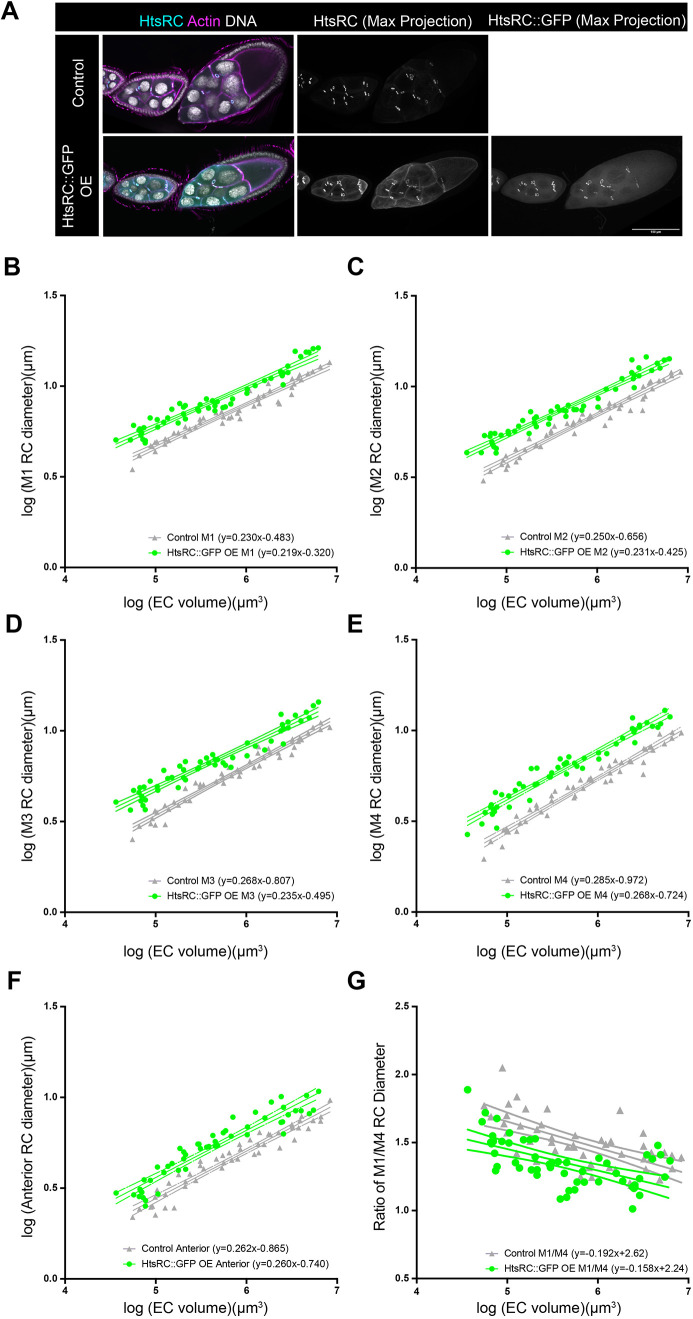
The size of subcellular structures must be tightly controlled to maintain normal cell function. Despite its importance, few studies have determined how the size of organelles or other structures is maintained during development, when cells are growing, dividing and rearranging. The developing Drosophila egg chamber is a powerful model in which to study the relative growth rates of subcellular structures. The egg chamber contains a cluster of 16 germline cells, which are connected through intercellular bridges called ring canals. As the egg chamber grows, the germline cells and the ring canals that connect them increase in size. Here, we demonstrate that ring canal size scaling is related to lineage; the largest, 'first-born' ring canals increase in size at a relatively slower rate than ring canals derived from subsequent mitotic divisions. This lineage-based scaling relationship is maintained even if directed transport is reduced, ring canal size is altered, or in egg chambers with twice as many germline cells. Analysis of lines that produce larger or smaller mature eggs reveals that different strategies could be used to alter final egg size.

Keywords: Drosophila; Intercellular bridge; Oogenesis; Ring canal.


Conflict of interest statement

Competing interests The authors declare no competing or financial interests.

Figures

Fig. 1.
Fig. 1.
Ring canal size and nuclear size vary within the egg chamber. (A) Single-plane confocal images of stage 5 (I), early stage 9 (II), and stage 10 (III) control egg chambers (w1118/+;;matαTub-GAL4) stained with an HtsRC antibody, phalloidin and DAPI. Lower panels show anterior (Ant), M4 and M1 ring canals from each of the indicated egg chambers (which were located in different z-planes than that shown in the upper panels); ring canal identity is indicated by the colored bars, which match those used in E. Measurements of these ring canals and associated nuclei are included below; measurements were made in the z-plane in which each structure was in focus. Scale bars: 50 µm (top); 5 µm (bottom). (B-D) Log-log plots of egg chamber (EC) volume, nuclear area, or ring canal (RC) diameter from stage 5-10b. All four posterior nuclei and ring canals (M1, M2, M3 and M4) were measured. Error bars in B,C are s.d. n=61 egg chambers. 95% confidence intervals are shown. (E) Diagram showing the stereotypical connections that result from the four rounds of mitotic divisions. Fill colors correspond to those used in D, and the thickness of the outlines and connecting lines correspond to distance from the oocyte (thicker lines are closer to the oocyte, and thinner lines are further from the oocyte).
Fig. 2.
Fig. 2.
Reducing directed transport and oocyte growth has a modest effect on ring canal size and scaling. (A) Single plane confocal images of control and dhc64C-RNAi egg chambers (otu-GAL4,UAS-Dcr-2) stained with an HtsRC antibody, phalloidin and DAPI. Scale bar: 50 µm. (B,C) Log-log plots of egg chamber (EC) volume, oocyte area, or ring canal (RC) diameter from stage 5-10b. 95% confidence intervals are shown. Error bars in C are s.d. (D) Scaling exponent for ring canals separated by lineage. Error bars are 95% confidence intervals. n=49 egg chambers for control and 28 egg chambers for dhc64C-RNAi.
Fig. 3.
Fig. 3.
Over-expression of HtsRC::GFP increases ring canal size without affecting the lineage-based scaling. (A) Single-plane or maximum intensity projections of confocal images of control and HtsRC::GFP over-expressing (OE) egg chambers (matαTub-GAL4) stained with an HtsRC antibody, phalloidin and DAPI. Scale bar: 100 µm. (B-F) Log-log plots of egg chamber (EC) volume and ring canal (RC) diameter. (G) The ratio of M1/M4 ring canal diameter from stage 5-10b. 95% confidence intervals are shown. n=50 egg chambers for control and 54 egg chambers for HtsRC::GFP OE.
Fig. 4.
Fig. 4.
Depletion of Tom6 increases the number of germline cells but does not alter the pattern of ring canal scaling. (A) Single-plane confocal images of control and tom6-RNAi egg chambers (nos-GAL4) stained with an HtsRC antibody, phalloidin and DAPI. Maximum intensity projections of the tom6-RNAi egg chambers are included to show the additional nurse cells and ring canals in these egg chambers. Scale bar: 50 µm. (B,C) Log-log plots of egg chamber (EC) volume and ring canal (RC) diameter from stage 5-10b. n=74 egg chambers for control and 64 egg chambers for tom6-RNAi. 95% confidence intervals are shown. (D) Scaling exponent for ring canals separated by lineage. Error bars indicate 95% confidence interval. Asterisks indicate significant difference between control and tom6-RNAi (ANCOVA, P<0.05).
Fig. 5.
Fig. 5.
Differences in posterior ring canal size and scaling in big egg and small egg D. melanogaster lines. (A) Box and whiskers plot of mature egg volume of big egg and small egg D. melanogaster lines. Median is indicated by the horizontal line, the box shows the 25-75th percentile, whiskers are the 10-90th percentile and points outside that range are shown. One-way ANOVA with Tukey's multiple comparisons test indicates mature eggs from all small egg lines (9.31.4, 8.26.2, 8.11.1, 7.17.4 and 9.2.2) are significantly smaller than all big egg lines (1.16.1, 2.15.4, 2.49.3, 1.40.2 and 3.34.1) (P<0.0001). n=20-77 eggs per line. (B) Scaling exponent for ring canals separated by lineage. Error bars indicate 95% confidence interval. There were significant differences in the scaling exponent across the four lines for M1, M2 and M4 (ANCOVA, P<0.05). (C-F) Log-log plots of egg chamber (EC) volume and ring canal (RC) diameter in two big egg (1.40.2 and 3.34.1) and two small egg (7.17.4 and 9.31.4) lines separated by lineage. 95% confidence intervals are shown. n=29 egg chambers for 1.40.2, 46 egg chambers for 3.34.1, 44 egg chambers for 7.17.4, and 43 egg chambers for 9.31.4.

Update of

References

    1. Ali-Murthy, Z., Fetter, R. D., Wang, W., Yang, B., Royer, L. A. and Kornberg, T. B. (2021). Elimination of nurse cell nuclei that shuttle into oocytes during oogenesis. J. Cell Biol. 220, e202012101. 10.1083/jcb.202012101 - DOI - PMC - PubMed
    1. Balachandra, S., Sarkar, S. and Amodeo, A. A. (2022). The nuclear-to-cytoplasmic ratio: coupling DNA content to cell size, cell cycle, and biosynthetic capacity. Annu. Rev. Genet. 56, 165-185. 10.1146/annurev-genet-080320-030537 - DOI - PMC - PubMed
    1. Cantwell, H. and Nurse, P. (2019). Unravelling nuclear size control. Curr. Genet. 65, 1281-1285. 10.1007/s00294-019-00999-3 - DOI - PMC - PubMed
    1. Carvalho, A., Desai, A. and Oegema, K. (2009). Structural memory in the contractile ring makes the duration of cytokinesis independent of cell size. Cell 137, 926-937. 10.1016/j.cell.2009.03.021 - DOI - PubMed
    1. Diegmiller, R., Doherty, C. A., Stern, T., Alsous, J. I. and Shvartsman, S. Y. (2021). Size scaling in collective cell growth. Development 148, dev.199663. 10.1242/dev.199663 - DOI - PMC - PubMed

LinkOut - more resources