. 2024 Aug 27;12(1):48.
doi: 10.1038/s41413-024-00354-4.

SREBP2 restricts osteoclast differentiation and activity by regulating IRF7 and limits inflammatory bone erosion

Affiliations

SREBP2 restricts osteoclast differentiation and activity by regulating IRF7 and limits inflammatory bone erosion

Haemin Kim et al. Bone Res. .

Abstract

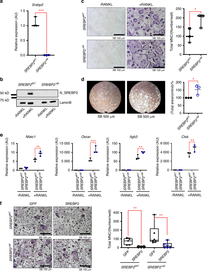
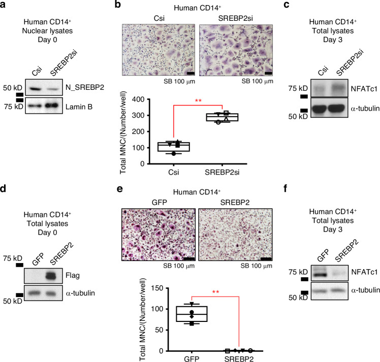
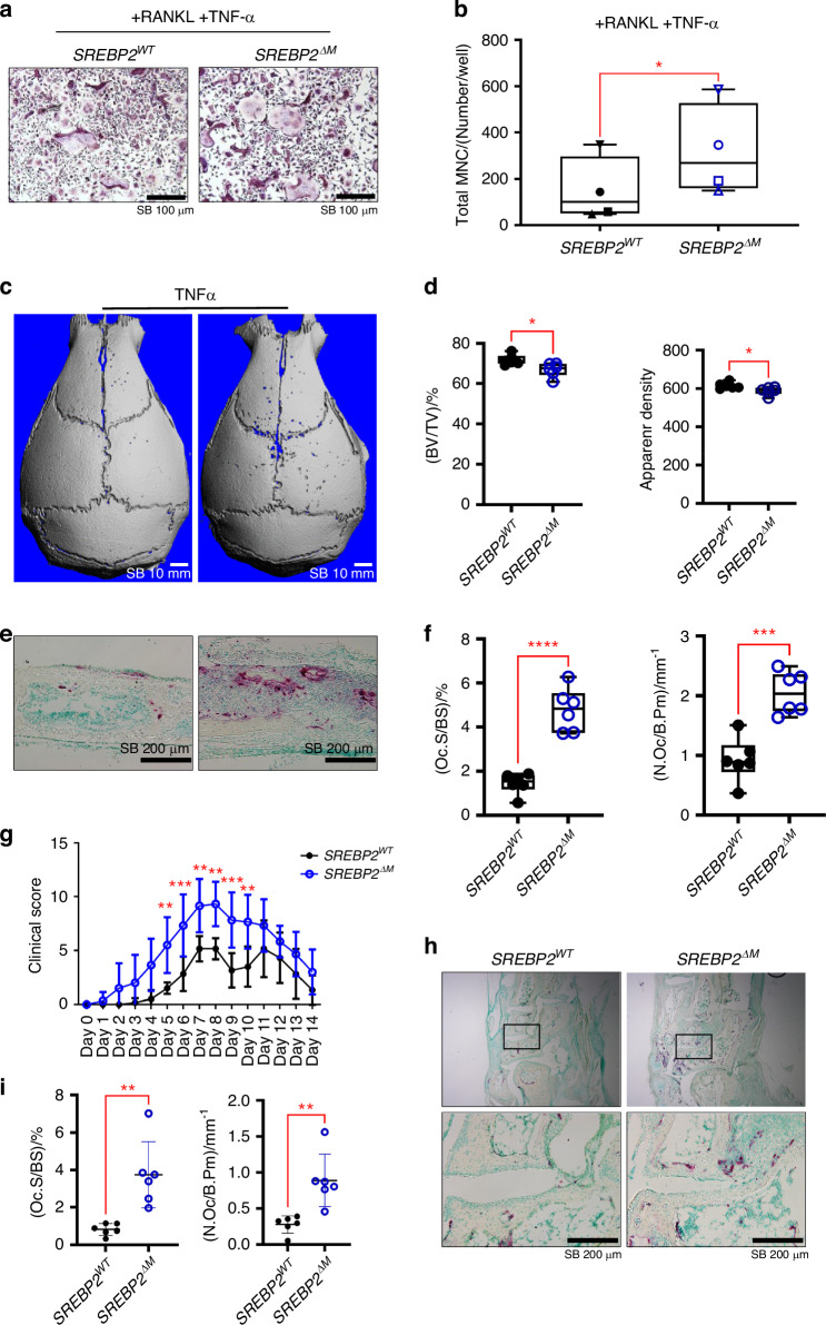
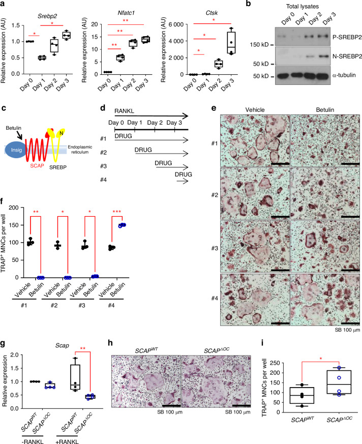
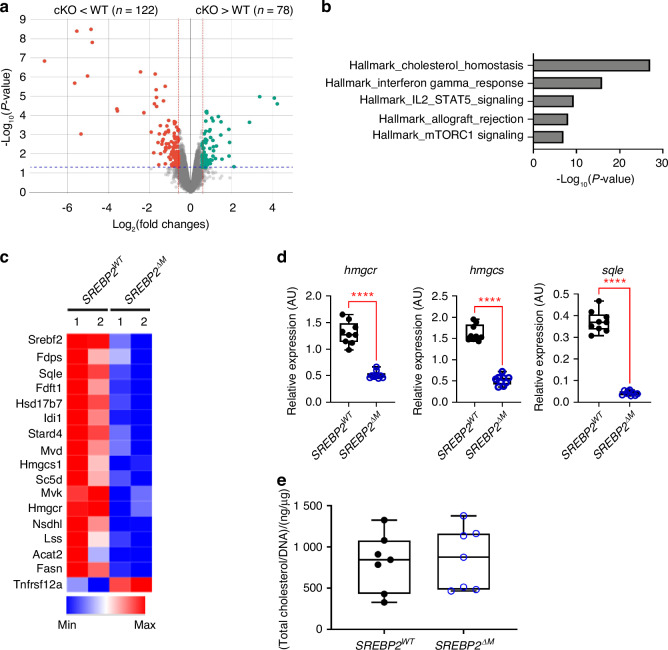
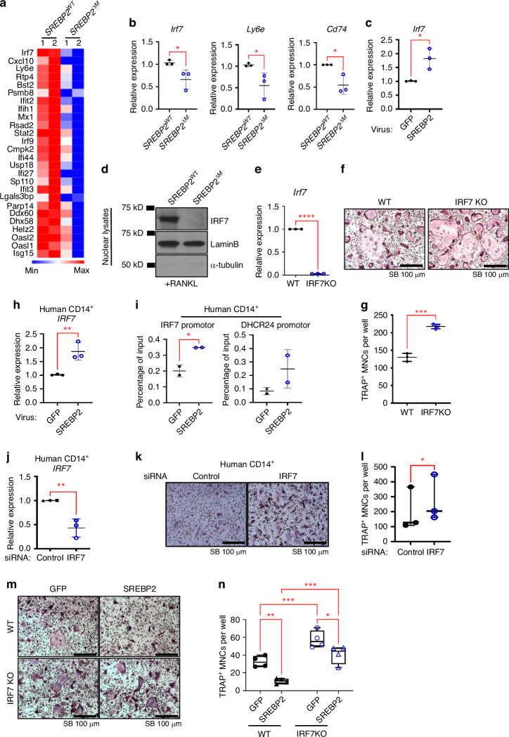
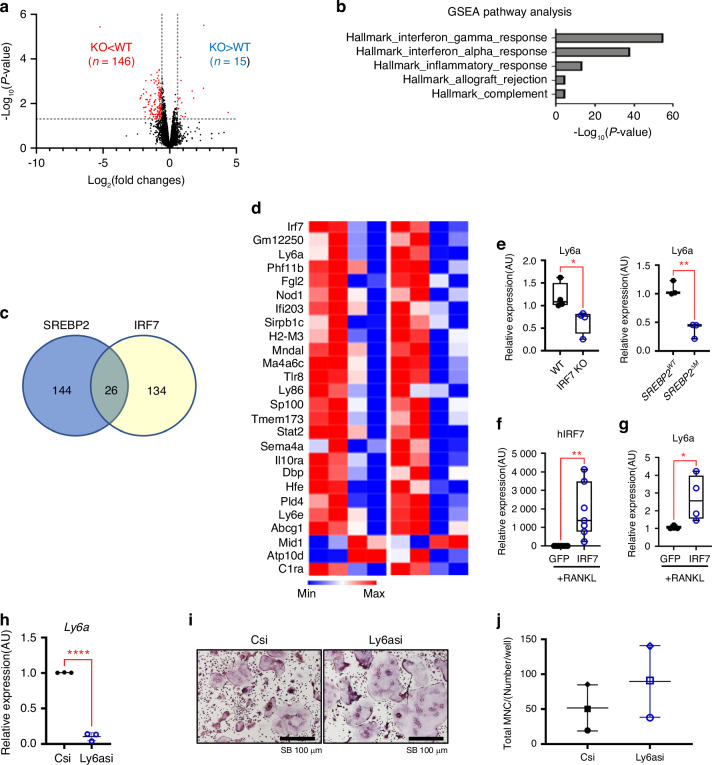
Osteoclasts are multinucleated bone-resorbing cells, and their formation is tightly regulated to prevent excessive bone loss. However, the mechanisms by which osteoclast formation is restricted remain incompletely determined. Here, we found that sterol regulatory element binding protein 2 (SREBP2) functions as a negative regulator of osteoclast formation and inflammatory bone loss. Cholesterols and SREBP2, a key transcription factor for cholesterol biosynthesis, increased in the late phase of osteoclastogenesis. The ablation of SREBP2 in myeloid cells resulted in increased in vivo and in vitro osteoclastogenesis, leading to low bone mass. Moreover, deletion of SREBP2 accelerated inflammatory bone destruction in murine inflammatory osteolysis and arthritis models. SREBP2-mediated regulation of osteoclastogenesis is independent of its canonical function in cholesterol biosynthesis but is mediated, in part, by its downstream target, interferon regulatory factor 7 (IRF7). Taken together, our study highlights a previously undescribed role of the SREBP2-IRF7 regulatory circuit as a negative feedback loop in osteoclast differentiation and represents a novel mechanism to restrain pathological bone destruction.


Conflict of interest statement

The authors declare no competing interests.

Figures

Fig. 1
Fig. 1
SREBP2 protects mice from bone loss. ad Femurs from 12-week-old female SREBP2WT and littermate control SREBP2∆M mice were subjected to microCT scan analysis. Representative 3D images of trabecular bone (a) or cortical bone (c) of distal femurs were shown. Scale bars: 1 mm. b Trabecular bone parameters of femurs from SREBP2WT (n = 10) and SREBP2∆M (n = 17) mice. Bone volume/ tissue volume (Tb. BV/TV), trabecular space (Tb. Sp), trabecular thickness (Tb.Th), and trabecular numbers (Tb.N). d Cortical bone parameters of SREBP2WT (n = 8) and SREBP2∆M (n = 11) mice. Bone volume per tissue volume (BV/TV) and porosity. e, f Histological assessment of femurs from 12-week-old female SREBP2 WT and SREBP2∆M mice (n = 8 per group). e Representative images. Decalcified sections of distal femurs of SREBP2 WT and SREBP2∆M mice were shown after TRAP staining. Methyl Green staining was followed to indicate bone. Scale bars: 200 µm. f The osteoclast surface per bone surface (Oc. S/BS), the number of osteoclasts per bone perimeter (N. Oc/B.Pm), eroded surface per bone surface (ES/BS) were quantified. g, h Bone dynamic labeling analysis from 12-week-old female SREBP2 WT and SREBP2∆M mice. g Representative images of calcein labeling. Scale bars: 200 µm. h Mineral apposition rate (MAR) and bone formation rate per bone surface (BFR/BS) (n = 6 per group). i The measurement of C-telopeptide of collagen type 1 (CTX) and Procollagen type I N-terminal propeptide (P1NP) from 12-week-old female SREBP2 WT and SREBP2∆M mice (n = 10 per group). All data are shown as median and interquartile range. n.s., not significant; **P < 0.01, ***P < 0.001, ****P < 0.000 1 by two-tailed unpaired t test
Fig. 2
Fig. 2
SREBP2 is a negative regulator of osteoclastogenesis. a Srebp2 mRNA levels were assessed by qPCR from SREBP2WT and SREBP2∆M BMMs. b–e SREBP2WT and SREBP2∆M BMMs cultured with RANKL (50 ng/mL) for three days. b Immunoblots of nuclear lysates using SREBP2 and Lamin B antibodies. c Cells were TRAP-stained (left) and counted for 3 or more nuclei (right). Scale bars: 100 µm. d Bone resorption activity wase assessed by culturing SREBP2WT and SREBP2∆M BMMs with RANKL (50 ng/mL). Scale bars: 500 µm. e Osteoclast marker genes were assessed after RANKL (50 ng/mL) stimulation for 3 days. f SREBP2WT and SREBP2∆M BMMs transduced with adenoviruses harboring GFP control or FLAG_N_SREBP2 (SREBP2) were cultured with RANKL (50 ng/mL) (left). Scale bars: 100 µm. TRAP-positive cells with 3 or more nuclei were counted (right). All data are shown as median and interquartile range. n.s., not significant; *P < 0.05; **P < 0.01; ***P < 0.001 by paired t test (a, c, d) or two-way ANOVA with multiple comparisons (e, f). All data are from at least 3 independent experiments
Fig. 3
Fig. 3
SREBP2 negatively regulates osteoclastogenesis in human CD14+ cells. ac Human CD14+ monocytes were nucleofected with control siRNA (Csi) or SREBP2-targeted siRNA (SREBP2si). a Immunoblots of nuclear lysates using SREBP2 and Lamin B antibodies before RANKL stimulation. b Nucleofected cells cultured with RANKL (40 ng/mL) were TRAP-stained (upper) and counted for 3 or more nuclei (lower). Scale bars: 100 µm. c Immunoblots of total lysates at day 3 of RANKL (40 ng/mL) stimulation using NFATc1 and α-tubulin antibodies. df Human CD14+ monocytes were transduced with adenoviral particles encoding GFP control (GFP) or FLAG-N-SREBP2 (N-SREBP2). d Immunoblots of total lysates using Flag and α-tubulin antibodies before RANKL stimulation. e Transduced cells cultured with RANKL (40 ng/mL) were TRAP-stained and counted for 3 or more nuclei. Scale bars: 100 µm. f Immunoblots of total lysates at day 3 of RANKL (40 ng/mL) stimulation using NFATc1 and α-tubulin antibodies. All data are shown as median and interquartile range. **P < 0.01 by paired t test (b, e). All data are from at least 3 independent experiments
Fig. 4
Fig. 4
SREBP2 protects mice from inflammatory bone destruction. a, b Osteoclastogenesis assay. SREBP2WT and SREBP2∆M BMMs were cultured with RANKL (20 ng/mL) plus TNF-α (20 ng/mL). a Representative images of TRAP staining. Scale bars: 100 µm. b Quantification of TRAP-positive cells with 3 or more nuclei. cf TNF-α-induced supracalvarial bone loss model (n = 6 per group). c Representative 3D images. d μCT analyses for bone volume/tissue volume (BV/TV) and apparent density. e Representative images for TRAP staining of histological sections of calvaria. Scale bars: 200 µm. f The osteoclast surface per bone surface (Oc. S/BS) and the number of osteoclasts per bone perimeter (N. Oc/B.Pm) were quantified. gi K/BxN serum-induced arthritis model (n = 6 per group). g Time course of clinical score. h Representative TRAP staining of histological sections of tarsal bones. Scale bars: 200 µm. i The osteoclast surface per bone surface (OcC. S/BS) and the number of osteoclasts per bone perimeter (N. Oc/B.Pm) were quantified. All data are shown as median and interquartile range. n.s., not significant; *P < 0.05; **P < 0.01; ***P < 0.005; ****P < 0.001 by two-tailed unpaired t test (b, d, f, i), or 2-way repeated measures ANOVA with multiple comparisons (g)
Fig. 5
Fig. 5
SCAP/SREBPs inactivation in late stage accelerates osteoclast differentiation. a Srebp2, Nfatc1 and Cathepsin K mRNA levels were assessed from 8-week-old female C57/BL6 mouse BMMs cultured with RANKL (50 ng/mL) for indicated days. b Immunoblots of total lysates from indicated days of culture after RANKL (50 ng/mL) stimulation using SREBP2 and α-tubulin antibodies. c Schematic showing the action of Betulin. d Schematic of drug treatment for experiments, #1 indicating drug treated since Day 0 of the culture (concurrent with RANKL), #2 from Day 1 of the culture, #3 from Day 2 of the culture, #4 from Day 3 of the culture. In case of #4, the drugs were incubated for 6 h. e, f 0 and 1 µg/mL of betulin were tested for four treatment schedules. e Representative images of TRAP-stained cells. f Numbers of TRAP-positive osteoclasts per well. g Scap mRNA levels were assessed by qPCR from SCAPWT and SCAP∆OC osteoclasts at day 3 of RANKL treatment. h Representative images of TRAP-stained cells. i Numbers of TRAP-positive osteoclasts from SCAPWT and SCAP∆OC are shown. All data are shown as median and interquartile range. n.s., not significant; *P < 0.05; **P < 0.01; ***P < 0.005; ****P < 0.001 by two-tailed unpaired t test (i), one-way (a) or two-way ANOVA with multiple comparisons (f, g). All data are from at least 3 independent experiments
Fig. 6
Fig. 6
Total mRNA sequencing reveals differentially regulated genes in SREBP2 deficient osteoclasts. a Volcano plot of RNA-sequencing analysis of differentially expressed genes. Red dots represent significantly down-regulated and green dots represent up-regulated genes (P < 0.05, fold changes >1.5). b Gene Set Enrichment Analysis (GSEA) for pathways are shown. c Heatmap of representative cholesterol homeostasis genes from SREBP2WT and SREBP2∆M BMMs stimulated with RANKL (50 ng/mL) for 3 days. d Hmgcr, Hmgcs, and Sqle mRNA levels were assessed from SREBP2WT and SREBP2∆M BMMs stimulated with RANKL (50 ng/mL) for 3 days. e SREBP2WT and SREBP2∆M BMMs stimulated with RANKL (50 ng/mL) for 3 days were subjected to intracellular cholesterol measurement. Relative intracellular cholesterols in SREBP2WT and SREBP2∆M osteoclasts measured by Amplex Red cholesterol assay kit (n = 7). All data are shown as median and interquartile range. n.s., not significant; ****P < 0.000 1 by two-tailed unpaired t test (d, e). All data are from two biological replicates (ac) and at least 3 independent experiments (d, e)
Fig. 7
Fig. 7
SREBP2 regulates IRF7. a Heatmap of representative interferon gamma response genes from SREBP2WT and SREBP2∆M BMMs stimulated with RANKL (50 ng/mL) for 3 days. b Irf7, Ly6e, and Cd74 mRNA levels were assessed from SREBP2WT and SREBP2∆M BMMs stimulated with RANKL (50 ng/mL) for 3 days. c Irf7 mRNA levels were assessed after adenoviral overexpression of GFP control or FLAG-N-SREBP2 (SREBP2) in mouse BMMs stimulated with RANKL (50 ng/mL). d Immunoblots of nuclear lysates from day 3 of RANKL (50 ng/mL) stimulation using anti-IRF7, SREBP2, Lamin B, and α-tubulin antibodies. e Irf7 mRNA levels were assessed from 8-week-old female wildtype and IRF7 knockout BMMs. f Representative images of TRAP staining of wildtype and IRF7 knockout BMMs cultured with RANKL (50 ng/mL). Scale bars: 100 µm. g Quantification of TRAP-positive cells with 3 or more nuclei. RAP-stained Wildtype and IRF7 knockout BMMs cultured with RANKL (50 ng/mL) were TRAP-stained (upper) and counted for 3 or more nuclei (lower). Scale bars: 100 µm. h IRF7 mRNA levels were assessed after adenoviral overexpression of GFP control or FLAG-N-SREBP2 (SREBP2) in human CD14+ monocytes stimulated with RANKL (40 ng/mL). i IRF7 and DHCR24 promoters were assessed by ChIP-qPCR analyses from human CD14+ monocytes transduced with adenoviruses harboring GFP control (GFP) or FLAG-N-SREBP2 (SREBP2). 2 independent experiments were performed from 4 different human donors. J–l IRF7 mRNA levels were assessed from human CD14+ monocytes nucleofected with control or IRF7 siRNAs (j). Human CD14+ monocytes nucleofected with control or IRF7 siRNAs cells were TRAP-stained after RANKL (40 ng/mL) stimulation (k) and counted for 3 or more nuclei (l). Scale bars: 100 µm. m–n Osteoclastogenesis assay. Wildtype and IRF7 knockout BMMs transduced with adenoviruses harboring GFP control or FLAG_N_SREBP2 (SREBP2) were cultured with RANKL (50 ng/mL). m Representative images were shown. Scale bars: 100 µm. n TRAP-positive cells with 3 or more nuclei were counted. All data are shown as median and interquartile range. n.s., not significant; *P < 0.05; **P < 0.01; ***P < 0.001; ****P < 0.000 1 by paired t test (b, c, e, g, h, i) or two-way ANOVA with multiple comparisons (n). All data are from two (a, i) and at least 3 independent experiments (bh)
Fig. 8
Fig. 8
The SREBP2-IRF7 axis regulates 26 overlapping genes. a Volcano plot of RNA-seq analysis of differentially expressed genes (DEGs). Blue dots represent significantly differentially regulated genes (P < 0.05, fold changes >1.5). b Gene Set Enrichment Analysis (GSEA) of DEG in 7 a. c Venn diagram showing genes commonly regulated by SREPB2 and IRF7. d Heatmap of overlapped genes in DEGs shown in 5 a and 7 a. e Ly6a mRNA levels were assessed in wildtype, IRF7 knockout, SREBP2WT, and SREBP2∆M BMMs after 3 days of RANKL (50 ng/mL) treatment. f IRF7 mRNA levels were assessed after adenoviral overexpression of GFP control or 3xHA_GFP_IRF7 (IRF7) in mouse BMMs stimulated with RANKL (50 ng/mL). g Ly6a mRNA levels were assessed after adenoviral overexpression of GFP control or 3xHA_GFP_IRF7 (IRF7) in mouse BMMs stimulated with RANKL (50 ng/mL). h Ly6a mRNA levels were assessed from control siRNAs (Csi) or ly6a siRNAs (Ly6asi) transfected BMMs. i Osteoclastogenesis assay. Csi or Ly6asi transfected BMMs were cultured with RANKL (50 ng/mL). Representative images were shown. Scale bars: 100 µm. j TRAP-positive cells with 3 or more nuclei were counted. All data are shown as median and interquartile range. n.s., not significant; *P < 0.05; **P < 0.01 by paired t-test (e,f,g,h,j). All data are from two biological replicates (ad) or at least 3 independent experiments (eh, j)

References

    1. Tsukasaki, M. & Takayanagi, H. Osteoimmunology: evolving concepts in bone-immune interactions in health and disease. Nat. Rev. Immunol.19, 626–642 (2019). 10.1038/s41577-019-0178-8 - DOI - PubMed
    1. Park-Min, K. H. Mechanisms involved in normal and pathological osteoclastogenesis. Cell Mol. Life Sci.75, 2519–2528 (2018). 10.1007/s00018-018-2817-9 - DOI - PMC - PubMed
    1. Teitelbaum, S. L. Bone resorption by osteoclasts. Science289, 1504–1508 (2000). 10.1126/science.289.5484.1504 - DOI - PubMed
    1. Udagawa, N. et al. Osteoprotegerin produced by osteoblasts is an important regulator in osteoclast development and function. Endocrinology141, 3478–3484 (2000). 10.1210/endo.141.9.7634 - DOI - PubMed
    1. Luo, J. et al. LGR4 is a receptor for RANKL and negatively regulates osteoclast differentiation and bone resorption. Nat. Med. 22, 539–546 (2016). 10.1038/nm.4076 - DOI - PubMed

MeSH terms

Substances