. 2024 Aug 27;15(1):7396.
doi: 10.1038/s41467-024-51686-y.

Lung megakaryocytes engulf inhaled airborne particles to promote intrapulmonary inflammation and extrapulmonary distribution

Affiliations

Lung megakaryocytes engulf inhaled airborne particles to promote intrapulmonary inflammation and extrapulmonary distribution

Jiahuang Qiu et al. Nat Commun. .

Abstract

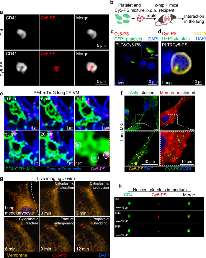
Many lung immune cells are known to respond to inhaled particulate matter. However, current known responses cannot explain how particles induce thrombosis in the lung and how they translocate to distant organs. Here, we demonstrate that lung megakaryocytes (MKs) in the alveolar and interstitial regions display location-determined characteristics and act as crucial responders to inhaled particles. They move rapidly to engulf particles and become activated with upregulation in inflammatory responses and thrombopoiesis. Comprehensive in vivo, in vitro and ex vivo results unraveled that MKs were involved in particle-induced lung damages and shed particle-containing platelets into blood circulation. Moreover, MK-derived platelets exhibited faster clotting, stronger adhesion than normal resting platelets, and inherited the engulfed particles from parent MKs to assist in extrapulmonary particle transportation. Our findings collectively highlight that the specific responses of MKs towards inhaled particles and their roles in facilitating the translocation of particles from the lungs to extrapulmonary organs for clearance.


Conflict of interest statement

The authors declare no competing interests.

Figures

Fig. 1
Fig. 1. Lung MKs accumulate more rapidly in the lung than other immune cells upon particle exposure.
a Number of AM, DC, NE, MO, MK, Baso and Eosin in the BALF increased at 6, 12, and 24 h hpi of CB particles (n = 4 mice per group). b Ratio of the cumulative increase of different cells (1-7) in BALF of treated mice in a relative to untreated control. Horizontal dotted line shows a ratio of 1. Ratio in each group is the mean values of 3 independent experiments. c Immunofluorescent images (left) and PF4+CD41+DAPI+ cell counts (right) show lung sections from mice post 24 h-exposure had more PF4+CD41+DAPI+ MKs (white arrow) than untreated control (n = 6 mice per group). d Fluorescent images of lung sections from naïve PF4-mTmG mice showing resident and crawling MKs (green) in the Alv. and Int. region (red, inside dotted line). e 3D fluorescent imaging of lung sections from untreated PF4-mTmG mice showing MK resident in Alv. wall. f Giemsa-Wright stained image of an isolated CD41+ Alv. MKs with the classical hyperchromatic nucleus and granular cytoplasm. g Fluorescent images of Alv. MKs (CD41+CD42b+ Hoechst+) obtained from BALF through magnetic bead sorting. h Fluorescence analysis of the percentage of Alv. MKs and Int. MKs in the whole lung from untreated PF4-mTmG mice (n = 3 mice per group). i Venn diagram showing the gene signature of Alv. and Int. MKs. Numbers represent number of genes. j Heatmap of the enriched pathways for genes expressed in Alv. and Int. MKs. k 3D fluorescent images showing accumulated PF4+GFP+MKs (green) and platelets (green) in lung sections from PF4-mTmG mice treated with CB particles for 24 h. l H&E and Masson stained images of lung sections from mice exposed to CB particles for 24 h show infiltration of inflammatory cells (blue arrows) and fiber deposition in alveoli (red arrows) and capillaries (green arrowheads). Data are presented as the mean values of 3 independent experiments ± SEM (a, c and h), P-value relative to 0 hpi (a) and control (c and h) by two-side student’s t-test.
Fig. 2
Fig. 2. Alv. and Int. MKs engulf particles.
a Schematic showing the interactions between fluorescently labeled Cy5-PSs (red) and Alv. or Int. MKs (green). b Imaging using two-photon intravital microscopy (2PIVM) shows lung MKs (green) migrating to engulf Cy5-PSs (pink) in PF4-mTmG mice treated with 2 mg/kg body weight Cy5-PSs through o.p.a. for 2 h. c Fluorescent images of Alv. MKs (green) crawling towards and engulfing Cy5-PSs (yellow arrows) deposited in the alveoli. Nuclei are stained with DAPI (blue). d Intravital two-photon imaging of lung MK (green) squeezing into and out of 2 bordering alveoli possibly through the pore of Kohn (white arrow). e Flow cytometry images show that CD41+ MKs (yellow) from the lungs of mice treated with Cy5-PSs for 12 h contain Cy5-PSs (red). Nuclei are stained with Hoechst (blue). f Fluorescent images showing engulfed Cy5-PSs (orange) inside CD41+ MKs sorted from mice and Dami cells treated with 200 ng/mL Cy5-PSs for 12 h. White dotted line outlines a cell. g TEM image of lung MKs treated with Cy5-PSs for 12 h. Red arrow in the insert image indicates engulfed particles. h Flow cytometer analysis of engulfed particles in lung MKs of mice treated with Cy5-PSs after 6 and 12 h (n = 10 mice per group). N.D. not detected. i Quantification of internalized Cy5-PSs in Alv. MKs, Int. MKs and AMs from mice in h after 6 h (n = 10 mice per group). Data are presented as the mean values of 3 independent experiments ± SEM (h, i), one-way analysis of variance (ANOVA). j, k RNA-seq analyses on Int. MKs (j) and Alv. MKs (k) show significant mRNA enrichment in thrombopoiesis and immune regulatory functions, respectively. The statistical analyses were performed using Metascape, one-side Fisher’s exact test (j, k). Figure 2a, created with BioRender.com, released under a Creative Commons Attribution-NonCommercial-NoDerivs 4.0 International license.
Fig. 3
Fig. 3. Lung MKs are activated for inflammatory responses.
ac RNA-seq analysis of CD41+ MKs from lung single-cell suspension obtained from mice treated with 2 mg/kg body weight CB particles for 24 h. The volcano plots show the Log2 fold change of gene expression in CB group relative to control group (Ctrl). a Volcano plot showing upregulated and downregulated genes in lung MKs, two-tailed Wilcoxon rank-sum test. b KEGG pathway enrichment analysis of annotated upregulated genes in CB particle-treated lung MKs were associated with thrombocytosis, immunity, and cell regulation signals, one-side Fisher’s exact test. c Heatmap showing differentially expressed genes in lung MKs after CB particle treatment, categorized by biological functions. Columns represent samples (n = 3). d, e RT-qPCR analyses of Caspase-1 (d) and IL-1β (e) mRNA expression in lung MKs sorted from mice treated with CB particles for 24 h (n = 6 biological replicates per group). f H&E-stained images of lung sections show that c-mpl-/- mice treated with CB particles for 24 h had fewer inflammatory foci (red arrows) than treated WT animals. g Flow cytometer analysis of lung single-cell suspension from animals in f (n = 6 per group). h ELISA assay of IL-1β content in sera and BALF from WT and c-mpl-/- mice upon CB particles for 24 h (n = 6 per group). i H&E (top) and Masson (bottom) stained images of lung sections from WT and c-mpl-/- mice at 72 h post-CB administration show accumulated inflammatory cells (red arrows) and collagen deposition (blue arrows). j Immunofluorescent images of vascular junction with CD31+ cells in lung sections from WT and c-mpl-/- mice at 72 h post-CB particle exposure. White arrowheads indicate damaged junctions. White dash-dots outline the vessel. Inset: magnified view of vascular junction. k, l Immunofluorescent images (k) and counts per field (l) of neutrophils (MPO+Ly6G+ cells, white arrowheads) in lung sections from WT and c-mpl-/- mice at 72 h after CB exposure (n = 6 mice per group). m Immunofluorescence-stained images of lung sections show that mice receiving MK transplant promoted inflammatory responses and recruited significantly more neutrophils than control mice without transplants. n ELISA assay of IL-1β content in sera of mice with (+) and without (-) MK transplant at 24 h post 2 mg/kg body weight CB exposure (n = 6 mice per group). o Schematic shows c-mpl-/- mice displayed greater lung damage and 1–2 days slower recovery from particle-triggered inflammation than WT mice. Box and whisker plots indicate median, interquartile range and 10th to 90th percentiles of the distribution (h, n). Data are presented as the mean values of 3 independent experiments ± SEM (d–e, g, h, l and n), two-side student’s t-test (d, e and g) and one-way ANOVA (h, l and n). N.S, not significant.
Fig. 4
Fig. 4. Inhaled particles activate MKs to release platelets into blood circulation.
a Fluorescent images of lung sections from PF4-mTmG mice treated with 2 mg/kg body weight Cy5-PSs for 24 h and untreated control. White arrow pointing to MKs with engulfed particles releasing platelets (white arrowhead). b Fluorescent image of lung MK releasing a nascent platelet (green arrow) into a Alexa Fluor 647-dextran dye -stained blood capillary (red arrow). c Number of resting and activated platelets in peripheral blood of CB particle-treated mice (n = 8). d Flow cytometer analysis plots of activated platelets (488SSC-405SSC+/lowCD41+CD62P+) in peripheral blood of control and CB particle-treated animals from c. e Scanning electron microscope images of resting and activated platelet from CB-treated animals. f Peripheral blood of mice (n = 8) treated with CB for 24 h clotted faster than untreated control. g Bright-field images and counts per field show more platelets (blue arrowheads) from platelet-rich plasma (PRP) of CB particle-treated mice attached on the collagen matrix than untreated control (n = 10 randomly captured images). h KEGG pathway enrichment analysis of upregulated genes associated with MK development and platelet production, one-side Fisher’s exact test. MKs were isolated from mice treated with CB particles for 24 h. i Ex vivo maturation assay involves culturing isolated CD41+ MKs in TPO/SCF/IL-6 medium. Fluorescent images of membrane-stained matured MKs and nascent platelets after 2 days. j Live-cell imaging of isolated MKs with engulfed Cy5-PSs using LSFM features an extended cytoplasm and membrane shedding nascent platelet clusters (white dotted lines). k, l Flow cytometer analysis of the number of total platelets (CD41+Hoechst-, k) and activated platelets (CD41+CD62p+Hoechst-, l) in culture medium. MKs were pretreated with 200 ng/mL Cy5-PSs and cultured for 2 days (n = 10 biological replicates). m Schematic showing that particle inhalation induces greater activated platelets in circulation than physiological conditions. Box and whisker plots indicate median, interquartile range and 10th to 90th percentiles of the distribution (f, g). Data are presented as the mean values of 3 independent experiments ± SEM (c, f, g, k and l), two-side student’s t-test (c, f, g, k and l). Figure 4i and m, created with BioRender.com, released under a Creative Commons Attribution-NonCommercial-NoDerivs 4.0 International license.
Fig. 5
Fig. 5. MK-derived platelets contain particles inherited from their parental MKs.
a Immunofluorescent images of CD41+ platelets from peripheral blood of mice exposed to 2 mg/kg body weight Cy5-PSs for 72 h show platelet colocalized with fluorescent particles (red). b Schematic shows o.p.a. transplantation of 1.0 ×10⁷ platelets and 50 ng Cy5-PS mixture into c-mpl-/- mice. c Fluorescent images of liver sections from recipient c-mpl-/- mice in b 72 h after transplantation showing Cy5-PSs adhered onto platelets (Green). d Fluorescent images of lung sections in recipient mice showing platelets (green) and Cy5-PSs (red) inside CD45+ immune cells (yellow) at 72 h post platelets-Cy5-PS mixture transplantation. e Intravital two-photon imaging shows lung MK (green) releasing nascent platelets containing Cy5-PSs (pink). Red dotted square is magnified in bottom right. f Fluorescent images of Dami cells treated with 200 ng/mL Cy5-PSs for 12 h showing endoplasmic distribution of engulfed particles. Phalloidin-stained cytoskeleton (green, left panel), carbocyanine-stained membrane (red, right panel) and engulfed fluorescent particles. g Continuous imaging analysis of MKs every 3 min. Nascent platelet clusters released from Cy3-labeled MKs (yellow) are shown. h Flow cytometry images of Cy5-PSs (red)-laden platelets (CD41+, green) collected from MK induction medium. Figure 5b, created with BioRender.com, released under a Creative Commons Attribution-NonCommercial-NoDerivs 4.0 International license.
Fig. 6
Fig. 6. Extrapulmonary transportation of inhaled particles through MK-derived platelets.
a Schematic shows o.p.a. transplantation of particle-laden CD41+GFP+ MKs into c-mpl-/- mice for tracking platelet distribution. b Fluorescent images of lung sections from recipient c-mpl-/- mice show MKs (CD41+GFP+DAPI+) remain loaded with Cy5-PSs (red) 72 h after transplantation. c Immunofluorescent images of liver sections from recipient c-mpl-/- mice 72 h after MK transplantation. Donor MK-derived platelets (green) overlap with Cy5-PSs (red). d Ratio of platelet-bound and platelet-free particles in liver, spleen and kidney determined by RFI analysis of fluorescent images (n = 6 randomly captured images). e Fluorescent images show donor-derived platelet (green) in the liver is transported through blood vessel (Alexa Fluro 647-dextran+, circled out with white dotted line). f Immunofluorescent images of EVs (CD63+, yellow) and Cy5-PS (red) in liver sections from recipient c-mpl-/-mice 72 h after MK transplantation. CD41+GFP+ donor MK-derived platelets (green) with engulfed Cy5-PSs (red) are shown in the bottom as reference. g Fluorescent images of liver sections from WT and c-mpl-/- mice after exposure to Cy5-PSs for 72 h. Fewer particles are seen in c-mpl-/- mice. h ICP-OES analysis of SiO2 content in the lung, liver, kidney, spleen and peripheral blood of WT and c-mpl-/- mice treated with 2 mg/kg body weight SiO2 particles for 72 h (n = 8 mice per group). i Regression analysis of the particle content in the liver based on RFI quantification and ICP-OES method. Dotted lines represent 95% confidence intervals for the linear regression line. Each dot corresponds to one individual mouse sample. Statistical analysis was performed by linear regression analysis, two-side Pearson correlation test. j Counts per field of TUNEL positive cells in liver sections from WT and c-mpl-/- mice at 24 and 72 h after CB exposure (n = 3 mice per group). Box and whisker plots indicate median, interquartile range and 10th to 90th percentiles of the distribution (d, h). Data are presented as the mean values of 3 independent experiments ± SEM (d, h and j), P-value by one-way ANOVA (d, h and j). N.S, not significant. Figure 6a, created with BioRender.com, released under a Creative Commons Attribution-NonCommercial-NoDerivs 4.0 International license.
Fig. 7
Fig. 7
Schematic showing extrapulmonary transport of inhaled particles through the MK-platelet axis.

Similar articles

Cited by

References

    1. Weichenthal, S. et al. Association of sulfur, transition metals, and the oxidative potential of outdoor PM2.5 with acute cardiovascular events: a case-crossover study of Canadian adults. Environ. Health Perspect.129, 107005 (2021). 10.1289/EHP9449 - DOI - PMC - PubMed
    1. Shah, A. S. et al. Global association of air pollution and heart failure: a systematic review and meta-analysis. Lancet382, 1039–1048 (2013). 10.1016/S0140-6736(13)60898-3 - DOI - PMC - PubMed
    1. Li, D. et al. Fluorescent reconstitution on deposition of PM(2.5) in lung and extrapulmonary organs. Proc. Natl Acad. Sci. USA116, 2488–2493 (2019). 10.1073/pnas.1818134116 - DOI - PMC - PubMed
    1. Choi, H. S. et al. Rapid translocation of nanoparticles from the lung airspaces to the body. Nat. Biotechnol.28, 1300–1303 (2010). 10.1038/nbt.1696 - DOI - PMC - PubMed
    1. Liu, X. et al. Serum apolipoprotein A-I depletion is causative to silica nanoparticles-induced cardiovascular damage. Proc. Natl Acad. Sci. USA118, e2108131118 (2021). 10.1073/pnas.2108131118 - DOI - PMC - PubMed

Substances

LinkOut - more resources