. 2024 Aug 27;24(1):59.
doi: 10.1186/s12896-024-00885-y.

Purification and characterization of soluble recombinant Crimean-Congo hemorrhagic fever virus glycoprotein Gc expressed in mammalian 293F cells

Affiliations

Purification and characterization of soluble recombinant Crimean-Congo hemorrhagic fever virus glycoprotein Gc expressed in mammalian 293F cells

Nigel Aminake Makoah et al. BMC Biotechnol. .

Abstract

Background: Crimean-Congo hemorrhagic fever (CCHF) is a tick-borne zoonotic disease that presents with severe hemorrhagic manifestations and is associated with significant fatality rates. The causative agent, Crimean-Congo Hemorrhagic Fever Virus (CCHFV), is a high-priority pathogen identified by the World Health Organization with no approved vaccine or specific treatment available. In addition, there is a critical need for enhanced diagnostic tools to improve public health awareness, prevention measures, and disease control strategies.

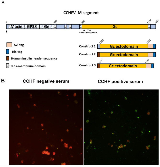
Methods: We designed plasmids to enable the purification of soluble CCHFV glycoprotein Gc expressed in mammalian 293 F cells, followed by purification using affinity and size exclusion chromatography. The purified antigen was analyzed by SDS-PAGE and Western blotting to confirm its reactivity to antibodies from CCHF survivors. Additionally, an in-house indirect ELISA was developed using the purified Gc as a coating antigen.

Results: The optimized expression system successfully produced soluble and pure Gc antigen after affinity chromatography. The protein showed specific reactivity with CCHFV-positive serum antibodies in Western blot analysis. The indirect ELISA assay demonstrated high efficacy in distinguishing between CCHFV-positive and -negative serum samples, indicating its potential as a valuable diagnostic tool. Size exclusion chromatography further confirmed the presence of aggregates in our protein preparation.

Conclusions: The purified Gc antigen shows promise for developing direct diagnostic assays for CCHFV. The antigen's suitability for subunit vaccine development and its application as bait for monoclonal antibody isolation from survivors could be investigated further. This work lays the foundation for future research into the development of rapid diagnostic tests for field deployment.

Keywords: CCHFV; Chromatography; Diagnostic; Expression; Glycoproteins; Purification.


Conflict of interest statement

The authors declare no competing interests.

Figures

Fig. 1
Fig. 1
A. Schematic depicting the M-segment of SPU 187/90 (top) encoding the mucin-like domain GP38, Gn, and Gc and transmembrane domains. Below are the constructs for recombinant protein expression (bottom right). Numbers correspond to full-length glycoprotein amino acid precursor numbering. Ectodomains, transmembrane (TM) domains, and endodomains were predicted using the TMHMM server v. 2.0. B. Localization of expressed CCHFV Gc by immunofluorescence assay: HEK293 cells transfected with construct 1 were stained with the human anti-CCHFV serum sample followed by the goat anti-human IgG (H + L) antibody conjugated with FITC. A negative CCHF serum was used as a control
Fig. 2
Fig. 2
A. Schematic of the plasmid: Gc-ecto(Ins) represents construct 3 in Fig. 1A. B-C. Analysis of CCHFV Gc under non-reducing conditions: Supernatant from construct 3 transfected 293 F cells was passed through a nickel agarose column and the eluted protein was analyzed by SDS-PAGE (B) and western blotting (C). D-E: Analysis of CCHFV Gc under reducing conditions. Supernatant from construct 3 transfected 293 F cells was passed through a nickel agarose column, the eluted protein was treated with the reducing agent DTT and analyzed by SDS-PAGE (D) and western blotting (E). F: Analysis of CCHFV Gc by Native PAGE. Supernatant from construct 3 transfected 293 F cells was passed through a nickel agarose column, the eluted protein was treated with 1% triton X-100 and resolved on a 4–12% native PAGE gel. PageRuler™ Pestained Protein Ladder (#26616;ThermoFisher Scientific, UK) was used to estimate the size of the protein on the SDS-PAGE gels and Western blot membranes
Fig. 3
Fig. 3
A. SEC elution profiles of CCHFV Gc: Gc protein purified by affinity chromatography was concentrated and stored at -80⁰C. Subsequently thawed and loaded on a AKTA™ Go and separated using a Superdex 200 increase 10/300 GL column. The stored solution is composed of aggregates (peak 1), multimers (peak?) and monomers (peak 2). B. SEC elution profiles of Gel filtration standard: chromatogram of Gel Filtration Standard (Bio Rad cat# 151–1901) comprised of five proteins. The profile denotes five peaks of which molecular weights can be determined. From left to right, peak A: thyroglobulin (670 kDa), peak B: gamma globulin (158 kDa), peak C: ovalbumin (44 kDa), peak D: myoglobin (17 kDa), peak E: Vitamin B12 (1.35 kDa). C. Analysis of CCHFV Gc under non-reducing conditions by SDS-PAGE: Approximately 4 µg of peak 1 and peak 2 were loaded on the gel and separated as described in the method. D. Analysis of CCHFV Gc under non-reducing conditions by Western blot: Approximately 4 µg of peak 1 and peak 2 were loaded on the gel, separated, and blotted as described in the method. The SARS-CoV S-glycoprotein tagged with Avi- and Histidine using the same approach described for CCHFV Gc was used as a control in the experiment. PageRuler™ Pestained Protein Ladder (#26616;ThermoFisher Scientific, UK) was used to estimate the size of the protein on the SDS-PAGE gels and Western blot membranes
Fig. 4
Fig. 4
Reactivity of controls and CCHF positive sera to CCHFV Gc: A. ELISA curves showing sera reactivity of CCHFV Gc to CCHF-positive and CCHF-negative serum samples. B. Area under the curve (AUC) of positive and negative serum samples
Fig. 5
Fig. 5
A. Native PAGE: Analysis of CCHFV Gc (Peak 1 and 2) by Native PAGE. Approximately 10 µg of the protein obtained from SEC was treated with 1% triton X-100 and resolved on a 4–12% native PAGE gel. B. ELISA: curves showing sera reactivity of CCHFV Gc monomer (blue) and aggregates (black) to CCHF-positive (66/08) and CCHF-negative (49/13) serum samples

References

    1. Hawman DW, Feldmann H. Crimean–Congo haemorrhagic fever virus. Nat Rev Microbiol. 2023;21:463–77. 10.1038/s41579-023-00871-9 - DOI - PMC - PubMed
    1. Ergönül Ö. Crimean-Congo haemorrhagic fever. Lancet Infect Dis. 2006;6:203–14. 10.1016/S1473-3099(06)70435-2 - DOI - PMC - PubMed
    1. Bente DA, Forrester NL, Watts DM, McAuley AJ, Whitehouse CA, Bray M. Crimean-Congo hemorrhagic fever: history, epidemiology, pathogenesis, clinical syndrome and genetic diversity. Antiviral Res. 2013;100:159–89. 10.1016/j.antiviral.2013.07.006 - DOI - PubMed
    1. Shahhosseini N, Wong G, Babuadze G, Camp JV, Ergonul O, Kobinger GP, et al. Crimean-Congo Hemorrhagic Fever Virus in Asia, Africa and Europe. Microorganisms. 2021;9:1907. 10.3390/microorganisms9091907 - DOI - PMC - PubMed
    1. Msimang V, Weyer J, le Roux C, Kemp A, Burt FJ, Tempia S, et al. Risk factors associated with exposure to Crimean-Congo haemorrhagic fever virus in animal workers and cattle, and molecular detection in ticks, South Africa. PLoS Negl Trop Dis. 2021;15:e0009384. 10.1371/journal.pntd.0009384 - DOI - PMC - PubMed

Publication types

MeSH terms

LinkOut - more resources