Case Reports
. 2024 Jul 28;16(7):e65559.
doi: 10.7759/cureus.65559. eCollection 2024 Jul.

Enhancing Anterior Esthetic Zone Implant Placement Through Bone Manipulation Techniques: A Case Series

Affiliations
Case Reports

Enhancing Anterior Esthetic Zone Implant Placement Through Bone Manipulation Techniques: A Case Series

Indu Raj et al. Cureus. .

Abstract

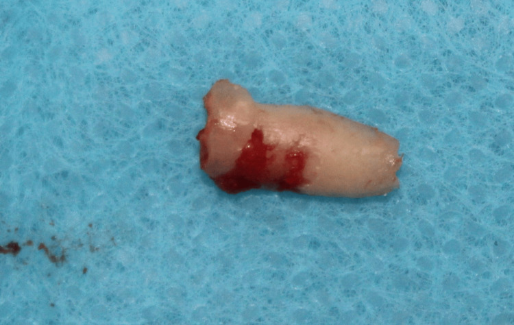
Replacement of missing teeth using dental implants has become the most frequently performed procedure. Following tooth loss, buccolingual bone width decreases significantly compromising the successful placement of dental implants. Various treatment modalities have been advocated in scenarios with insufficient buccolingual bone width like narrow diameter implant placement, horizontal guided bone regeneration, ridge splitting technique, and osseodensification. Maxillary anterior tooth loss is of prime esthetic concern, which needs immediate attention. Guided bone regeneration is the gold standard for patients presenting for rehabilitation in the anterior maxilla with inadequate buccolingual bone width. However, bone grafting techniques require longer treatment time; hence, various other techniques like lateral bone expansion, osseodensification, or socket shield technique are sought. This case series presents successful rehabilitation of the maxillary anterior esthetic zone with dental implants using various bone manipulation techniques, including lateral bone condensation, socket shield technique, and osseodensification.

Keywords: buccolingual width; expansion; implants; osseodensification; socket shield.


Conflict of interest statement

Human subjects: Consent was obtained or waived by all participants in this study. Conflicts of interest: In compliance with the ICMJE uniform disclosure form, all authors declare the following: Payment/services info: All authors have declared that no financial support was received from any organization for the submitted work. Financial relationships: All authors have declared that they have no financial relationships at present or within the previous three years with any organizations that might have an interest in the submitted work. Other relationships: All authors have declared that there are no other relationships or activities that could appear to have influenced the submitted work.

Figures

Figure 1
Figure 1. Case 1: Preoperative intra-oral image depicting narrow alveolar bone with buccal concavity and adequate soft tissue thickness in relation to missing 21
Figure 2
Figure 2. Case 1: Coronal section of CBCT image showing 3.13 mm of buccolingual bone at the crest, 3.69 mm bone at the middle third, and 4.77 mm bone apically denoting inadequate buccolingual bone for implant placement using conventional osteotomy
CBCT: Cone beam computed tomography.
Figure 3
Figure 3. Case 1: Bone expansion screw/thread former used for lateral bone expansion in narrow ridges or ridges with buccal concavity, which is used sequentially with increasing diameters from 1.6 to 2.8 mm and 1.9 to 3.4 mm
Figure 4
Figure 4. Case 1: Lateral bone expansion done using bone expansion screws of increasing diameters with the help of a ratchet that allows half of a turn at a time
Figure 5
Figure 5. Case 1: After assessment of successful osseointegration, the prosthetic phase was done, and the implant with respect to 21 was loaded using a cement-retained crown as the screw access hole was labial
Figure 6
Figure 6. Case 1: Intra-oral periapical radiograph was taken for the patient at the follow-up visit after one year, showing mild bone loss of 0.2 mm, which is the expected amount of bone loss according to Albrektsson's criteria
Figure 7
Figure 7. Case 2: Preoperative intra-oral image showing fractured 11 at the level of cementoenamel junction, with adequate soft tissue thickness but lacking the necessary ferrule for salvaging the tooth using post and core
Figure 8
Figure 8. Case 2: Preoperative RVG image showing root canal treated 21 fractured at CEJ
RVG: Radiovisiography; CEJ: Cementoenamel junction.
Figure 9
Figure 9. Case 2: Intra-oral image showing vertical sectioning of the root into buccal and palatal fragments using a long shank bur (Jull Dent-094A) of length 28 mm in a high-speed handpiece
Figure 10
Figure 10. Case 2: RVG taken intra-operatively to assess the level of root resection
RVG: Radiovisiography, digital x-ray.
Figure 11
Figure 11. Case 2: After vertical sectioning, the palatal fragment along with the apex of tooth 11 was extracted with the help of periotome to preserve the buccal root and buccal bone
Tooth number 11 = maxillary right central incisor.
Figure 12
Figure 12. Case 2: The remaining labial shield was reduced coronally to 1 mm above the alveolar crest using a round bur in a high-speed handpiece, and bevel preparation was done
Figure 13
Figure 13. Case 2: Osteotomy was then sequentially prepared, and a 3.5 x 13 mm implant was inserted palatal to the socket shield
Figure 14
Figure 14. Case 2: The jump gap between the implant and the shield was filled with 100 mg of osseograft (xenogenic particulate bone graft), which is a DMBM
DMBM: Demineralized bone matrix xenograft.
Figure 15
Figure 15. Case 2: Implant in relation to 11 was loaded after a period of eight months with a cement-retained crown
Figure 16
Figure 16. Case 2: During the follow-up visit one year after loading, IOPA was taken, which shows the intact buccal shield and a negligible amount of bone loss in relation to 11
IOPA: Intra-oral periapical radiograph.
Figure 17
Figure 17. Case 3: Preoperative intra-oral image showing narrow edentulous space with respect to missing maxillary left central incisor with an adequate soft tissue thickness
Figure 18
Figure 18. Case 3: The coronal section of the CBCT image of the patient showing 1.56 mm B-L width at the crest, 2.69 mm in the middle third, and 3.76 mm at the apical region of 21 region, which is inadequate for implant placement using conventional osteotomy
B-L: Buccolingual.
Figure 19
Figure 19. Case 3: Densah burs are used in counterclockwise rotation for the preparation of osteotomy to achieve bone condensation and improve primary stability
Figure 20
Figure 20. Case 3: Image depicting densah bur, which has more land than conventional straight drills that helps in controlled plastic deformation of bone
Figure 21
Figure 21. Case 3: Immediate postoperative IOPA depicting bone condensation with respect to 21
IOPA: Intra-oral periapical radiograph.
Figure 22
Figure 22. Case 3: Customized abutment was fabricated and torqued for 20 Ncm followed by luting of a cement-retained crown on the implant with respect to 21
Figure 23
Figure 23. Case 3: IOPA taken during follow-up visit after six months showing successful functioning of the prosthesis without bone loss
IOPA: Intra-oral periapical radiograph.

References

    1. Regeneration and enlargement of jaw bone using guided tissue regeneration. Buser D, Brägger U, Lang NP, Nyman S. Clin Oral Implants Res. 1990;1:22–32. - PubMed
    1. Horizontal ridge augmentation in conjunction with or prior to implant placement in the anterior maxilla: a systematic review. Kuchler U, von Arx T. Int J Oral Maxillofac Implants. 2014;29:14–24. - PubMed
    1. The alveolar ridge splitting/expansion technique: a systematic review. Bassetti MA, Bassetti RG, Bosshardt DD. Clin Oral Implants Res. 2016;27:310–324. - PubMed
    1. Lateral bone condensing and expansion for placement of endosseous dental implants: a new technique. Siddiqui AA, Sosovicka M. J Oral Implantol. 2006;32:87–94. - PubMed
    1. The socket-shield technique to support the buccofacial tissues at immediate implant placement. Gluckman H, Toit JD, Salama M. https://d1wqtxts1xzle7.cloudfront.net/45110043/gluckman-libre.pdf?146170... International Dentistry - African Edition. 2015;5:6–14.

Publication types

LinkOut - more resources