. 2024 Aug 27:7:0451.
doi: 10.34133/research.0451. eCollection 2024.

Circular RNA IGF1R Promotes Cardiac Repair via Activating β-Catenin Signaling by Interacting with DDX5 in Mice after Ischemic Insults

Affiliations

Circular RNA IGF1R Promotes Cardiac Repair via Activating β-Catenin Signaling by Interacting with DDX5 in Mice after Ischemic Insults

Tian-Kai Shan et al. Research (Wash D C). .

Abstract

The potential of circular RNAs (circRNAs) as biomarkers and therapeutic targets is becoming increasingly evident, yet their roles in cardiac regeneration and myocardial renewal remain largely unexplored. Here, we investigated the function of circIGF1R and related mechanisms in cardiac regeneration. Through analysis of circRNA sequencing data from neonatal and adult cardiomyocytes, circRNAs associated with regeneration were identified. Our data showed that circIGF1R expression was high in neonatal hearts, decreased with postnatal maturation, and up-regulated after cardiac injury. The elevation was validated in patients diagnosed with acute myocardial infarction (MI) within 1 week. In human induced pluripotent stem cell-derived cardiomyocytes (hiPSC-CMs) and myocardial tissue from mice after apical resection and MI, we observed that circIGF1R overexpression enhanced cardiomyocyte proliferation, reduced apoptosis, and mitigated cardiac dysfunction and fibrosis, while circIGF1R knockdown impeded endogenous cardiac renewal. Mechanistically, we identified circIGF1R binding proteins through circRNA precipitation followed by mass spectrometry. RNA pull-down Western blot and RNA immunoprecipitation demonstrated that circIGF1R directly interacted with DDX5 and augmented its protein level by suppressing ubiquitin-dependent degradation. This subsequently triggered the β-catenin signaling pathway, leading to the transcriptional activation of cyclin D1 and c-Myc. The roles of circIGF1R and DDX5 in cardiac regeneration were further substantiated through site-directed mutagenesis and rescue experiments. In conclusion, our study highlights the pivotal role of circIGF1R in facilitating heart regeneration and repair after ischemic insults. The circIGF1R/DDX5/β-catenin axis emerges as a novel therapeutic target for enhancing myocardial repair after MI, offering promising avenues for the development of regenerative therapies.


Conflict of interest statement

Competing interests: The authors declare that they have no competing interests.

Figures
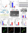
Fig. 1.
Fig. 1.
CircIGF1R is highly expressed in neonatal hearts and up-regulated in response to AR and MI model. (A) Volcano plot of the differentially expressed circRNAs between the neonatal and the adult rat hearts. (B) The expression level of rnoCirc_000455 is shown in RPM (reads per million mapped reads). (C) Species conservation analysis of the circIGF1R sequence using the UCSC Genome Browser and BLAST Browser. (D and E) The circular structure of circIGF1R was confirmed by amplification from gDNA and cDNA with both convergent and divergent primers using agarose gel electrophoresis, followed by Sanger sequencing. (F) CircIGF1R and IGF1R mRNA expression levels in CMs determined by PCR after RNase R treatment. (G) Relative expression of circIGF1R determined by qRT-PCR in hearts of mice at different ages, n = 4 in each group. (H and I) Relative expression of circIGF1R determined by qRT-PCR in hearts of mice after AR and MI, n = 4 in each group. (J) Relative expression of circIGF1R determined by qRT-PCR in the plasma of patients with or without AMI within 7 days, n = 30 in each group. (K) RNA-FISH assays of circIGF1R distribution in P1 CMs. Scale bars, 20 μm. Data are presented as mean ± SEM. *P ≤ 0.05, **P ≤ 0.01, and ***P ≤ 0.001.
Fig. 2.
Fig. 2.
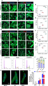
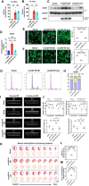
CircIGF1R mediates mice CM proliferation and apoptosis in vitro. (A to F) Representative pictures and quantification analysis of CM proliferation quantified by immunofluorescence for DNA synthesis (EdU), cell-cycle activity (Ki67), and mitosis (pH3) in P1 NMCMs transfected with Ad5: cTNT-sicircIGF1R, Ad5: cTNT-CON, and Ad5: cTNT-circIGF1R, n = 6 in each group. Scale bars, 50 and 12.5 μm. (G and H) Representative pictures and quantification analysis of CM apoptosis quantified by TUNEL staining in P1 NMCMs transfected with Ad5: cTNT-sicircIGF1R, Ad5: cTNT-CON, and Ad5: cTNT-circIGF1R after oxygen glucose deprivation (OGD), n = 6 in each group. Scale bars, 50 and 12.5 μm. (I and J) Cell flow cytometry was performed to detect the cell cycle of P1 NMCMs transfected with Ad5: cTNT-sicircIGF1R, Ad5: cTNT-CON, and Ad5: cTNT-circIGF1R, n = 3 in each group. (K and L) Representative pictures and quantification analysis of nucleation (mono-, bi-, and multi-) of primary P56 adult mouse cardiomyocytes (AMCMs) transfected with Ad5: cTNT-CON and Ad5: cTNT-circIGF1R, n = 3 in each group. Scale bar, 20 μm. Positive staining cardiomyocytes were indicated by arrows. Picture in each rectangular box was enlarged in the neighboring right panel. Data are presented as mean ± SEM. *P ≤ 0.05, **P ≤ 0.01, and ***P ≤ 0.001.
Fig. 3.
Fig. 3.
Overexpression of circIGF1R promotes human IPSC-CM proliferation. (A to H) Representative pictures and quantification analysis of CM proliferation quantified by immunofluorescence for DNA synthesis (EdU), cell-cycle activity (Ki67), mitosis (pH3), and cytokinesis (Aurora B) in human IPSC-CMs transfected with Ad5: cTNT-CON and Ad5: cTNT-circIGF1R, n = 6 in each group. Scale bars, 50 and 12.5 μm. (I and J) Cell flow cytometry was performed to detect the cell cycle of human IPSC-CMs transfected with Ad5: cTNT-CON and Ad5: cTNT-circIGF1R, n = 3 in each group. Positive staining cardiomyocytes were indicated by arrows. Picture in each rectangular box was enlarged in the neighboring right panel. Data are presented as mean ± SEM. *P ≤ 0.05, **P ≤ 0.01, and ***P ≤ 0.001.
Fig. 4.
Fig. 4.
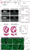
Knockdown of circIGF1R prevents neonatal mice cardiomyocyte proliferation following AR. (A) Experimental pattern: Ad5: cTNT-CON or Ad5:cTNT-sicircIGF1R was injected into myocardium following AR in P1 mice. Hearts were harvested at 6 and 22 days post-resection (dpr) to evaluate cardiomyocyte proliferation and scar area, respectively. Echocardiography was performed at 1 and 22 dpr to detect cardiac function. (B) Overall survival rate in mice treated with Ad5:cTNT-CON or Ad5:cTNT-sicircIGF1R, n = 20 in each group. (C and D) Cardiac function of ejection fraction and fractional shortening among the Ad5: cTNT-CON and Ad5: cTNT-sicircIGF1R treated mice at 1 and 22 day post-operation were detected by echocardiography, n = 6 in each group. (E and F) Masson’s trichrome staining was used to determine scar formation between the Ad5: cTNT-CON and Ad5: cTNT-sicircIGF1R treated mice at 22 dpr, n = 6 in each group. Scale bars, 500 μm and 300 μm. (G to J) Representative pictures and quantification analysis of CM proliferation quantified by cell-cycle activity (Ki67) and mitosis (pH3) in infarct border zone in Ad5: cTNT-CON and Ad5: cTNT-sicircIGF1R groups after AR, n = 6 in each group. Scale bars, 100 and 25 μm. Positive staining cardiomyocytes were indicated by arrows. Picture in each rectangular box was enlarged in the neighboring right panel. Data are presented as mean ± SEM. NS, no significance. *P ≤ 0.05, **P ≤ 0.01, and ***P ≤ 0.001.
Fig. 5.
Fig. 5.
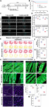
CircIGF1R promotes cardiac regeneration and repair in adult mice after MI. (A) Experimental pattern: AAV9: cTNT-CON or AAV9: cTNT-circIGF1R was injected into myocardium following MI in P56 mice. Hearts were harvested at 14 and 28 dpi to evaluate cardiomyocyte proliferation, apoptosis, and scar area, respectively. Echocardiography was performed at 1 and 28 dpi to detect cardiac function. (B) Overall survival rate in mice treated with AAV9: cTNT-CON or AAV9: cTNT-circIGF1R, n = 20 in each group. (C and D) Cardiac function of ejection fraction and fractional shortening among the sham-, AAV9: cTNT-CON-, or AAV9: cTNT-circIGF1R-treated mice at 1 and 28 day post-operation were detected by echocardiography, n = 5 in each group. (E to G) Masson’s trichrome staining and Sirius red staining were used to determine scar formation between the AAV9: cTNT-CON- or AAV9: cTNT-circIGF1R-treated mice at 28 dpi, n = 5 in each group. Scale bar, 1 mm. (H and I) Representative pictures and quantification analysis of CM proliferation quantified by DNA synthesis (EdU) in the infarct border zone in AAV9: cTNT-CON or AAV9: cTNT-circIGF1R groups after MI, n = 6 in each group. (J and K) Representative pictures and quantification analysis of CM apoptosis quantified by TUNEL staining in the infarct border zone in AAV9: cTNT-CON or AAV9: cTNT-circIGF1R groups after MI, n = 6 in each group. (L and M) Representative pictures and quantification analysis of CM size quantified by WGA immunofluorescence in the infarct border zone in AAV9: cTNT-CON or AAV9: cTNT-circIGF1R groups after MI, n = 6 in each group. Scale bars, 100 and 25 μm. Positive staining cardiomyocytes were indicated by arrows. Picture in each rectangular box was enlarged in the neighboring right panel. Data are presented as mean ± SEM. NS, no significance. *P ≤ 0.05, **P ≤ 0.01, and ***P ≤ 0.001.
Fig. 6.
Fig. 6.
CircIGF1R physically interacts with DDX5 protein and blocks its degradation via the ubiquitin–proteasomal pathway. (A) Proteins immunoprecipitated by the specific probe targeting the circIGF1R back-splice site were detected by silver staining. (B) Venn diagram of proteins interacting with circIGF1R, as identified by RNA pull-down assay and LC-MS/MS. (C) DDX5-specific peptide sequence identified using LC-MS/MS. (D) Western blot analysis following the RNA pull-down assay revealed the interaction between circIGF1R and DDX5, n = 3 in each group. (E) RIP assays confirmed that the DDX5 protein interacts with circIGF1R in CMs, n = 3 in each group. (F) Relative expression of circIGF1R determined by qRT-PCR in NMCMs transfected with Ad5: cTNT-CON and Ad5: cTNT-shDDX5. n = 6 in each group. (G) Prediction of circIGF1R-DDX5 interaction using the catRAPID algorithm. (H) Western blot analysis following the RNA pull-down assay verified the binding sequence of circIGF1R by using 2 biotinylated probes (151 to 202 and 426 to 477). (I) Visualized spatial structure of DDX5 protein using AlphaFold with functional protein domains. (J) Diagrams of full-length (FL) DDX5 proteins and truncations with domain depletion. (K) Binding domain of DDX5 for circIGF1R identified by an RIP assay. (L) qRT-PCR assays determining the relative mRNA expression levels of DDX5 after circIGF1R overexpression or knockdown. n = 3 in each group. (M and N) Western blot analysis determining the relative protein expression levels of DDX5 after circIGF1R overexpression or knockdown. n = 4 in each group. (O) Western blot analysis determining the expression of DDX5 in circIGF1R-overexpressed NMCMs treated with CHX (50 μg/ml). n = 3 in each group. (P and Q) Western blot analysis determining the expression of DDX5 in circIGF1R-silenced NMCMs treated with MG132 or CQ. n = 3 in each group. (R) Western blot analysis following Co-IP assay of the ubiquitination levels of DDX5 after circIGF1R overexpression. Data are presented as mean ± SEM. NS, no significance. *P ≤ 0.05, **P ≤ 0.01, and ***P ≤ 0.001.
Fig. 7.
Fig. 7.
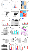
CircIGF1R up-regulates β-catenin signaling pathway and promotes transcriptional activation of c-Myc/Cyclin D1. (A) PCA was conducted to illustrate a distinct profile of circIGF1R overexpression in CMs compared to CMs treated with Ad5:cTNT-CON. (B) The volcano plot resulting from the quantitative transcriptomic analysis displays up-regulated (red) and down-regulated (blue) genes. Genes were considered significant if fold change >1.5 or <0.67, and FDR <0.05. (C) Cluster analysis and heatmap of DEGs. (D) Significant enriched GO biological process related to cardiac regeneration. (E) Significant enriched GO biological process related to Wnt signaling pathway. (F) Heatmap of DEGs associated with Wnt signaling pathway. (G) qRT-PCR assays determining the relative mRNA expression levels of β-catenin, cyclin D1, and c-Myc after circIGF1R overexpression. n = 3 in each group. (H and I) Relative expression of DDX5 and key proteins of β-catenin signaling pathway determined by Western blot in P1 NMCMs transfected with Ad5: cTNT-CON and Ad5: cTNT-circIGF1R, n = 3 in each group. (J and K) Relative expression of DDX5 and key proteins of β-catenin signaling pathway determined by Western blot in P1 NMCMs transfected with Ad5: cTNT-CON, Ad5: cTNT-circIGF1R, and Ad5: cTNT-circIGF1R + Ad5: cTNT-shDDX5, n = 3 in each group. (L) Graphical representation of 3-dimensional structures of the DDX5–β-catenin complex. Green: DDX5; purple: β-catenin. (M and N) Co-IP assays were carried out to confirm the interaction between DDX5 and β-catenin in P1 NMCMs. (O and P) CHIP assay to analyze the enrichment of cyclin D1 and c-Myc by β-catenin antibody, n = 6 in each group. (Q) CHIP assay to analyze the enrichment of cyclin D1 and c-Myc in P1 NMCMs transfected with Ad5: cTNT-CON and Ad5: cTNT-circIGF1R, n = 6 in each group. (R) TOP/FOP flash reporter assay in P1 NMCMs transfected with Ad5: cTNT-CON and Ad5: cTNT-circIGF1R, n = 6 in each group. Data are presented as mean ± SEM. NS, no significance. *P ≤ 0.05, **P ≤ 0.01, and ***P ≤ 0.001.
Fig. 8.
Fig. 8.
Blocking the DDX5 binding abolishes the role of circIGFlR in promoting cardiomyocytes proliferation and cardiac repair after MI. (A and B) Relative expression of circIGF1R in P1 NMCMs transfected with Ad5: cTNT-CON, Ad5: cTNT-circIGF1R, and Ad5: cTNT-circIGF1R mut and MI hearts transfected with AAV9: cTNT-CON, AAV9: cTNT-circIGF1R, and AAV9: cTNT-circIGF1R mut determined by qRT-PCR targeting the back-splicing site of circIGF1R, n = 6 in each group. (C and D) Relative expression of DDX5 protein determined by RNA pull-down and Western blot in P1 NMCMs transfected with Ad5: cTNT-CON, Ad5: cTNT-circIGF1R, and Ad5: cTNT-circIGF1R mut, n = 3 in each group. (E) Representative pictures and quantification analysis of CM proliferation quantified by immunofluorescence for DNA synthesis (EdU) in P1 NMCMs transfected with Ad5: cTNT-CON, Ad5: cTNT-circIGF1R, and Ad5: cTNT-circIGF1R mut, n = 6 in each group. Scale bar, 50 μm. (F) Representative pictures and quantification analysis of CM apoptosis quantified by TUNEL staining in P1 NMCMs transfected with Ad5: cTNT-CON, Ad5: cTNT-circIGF1R, and Ad5: cTNT-circIGF1R mut after OGD, n = 6 in each group. Scale bar, 50 μm. (G and H) Cell flow cytometry was performed to detect the cell cycle of P1 NMCMs transfected with Ad5: cTNT-CON, Ad5: cTNT-circIGF1R, and Ad5: cTNT-circIGF1R mut, n = 3 in each group. (I and J) Cardiac function of ejection fraction and fractional shortening among the sham group, vector group, circIGF1R group, and circIGF1R mut group at 1 and 28 day post-operation were detected by echocardiography, n = 5 in each group. (K to M) Masson staining and Sirius red staining were used to determine scar formation among the AAV9: cTNT-CON, AAV9: cTNT-circIGF1R, and AAV9: cTNT-circIGF1R mut at 28 dpi, n = 4 in each group. Scale bar, 1 mm. Data are presented as mean ± SEM. NS, no significance. *P ≤ 0.05, **P ≤ 0.01, and ***P ≤ 0.001.

References

    1. Ambrosy AP, Fonarow GC, Butler J, Chioncel O, Greene SJ, Vaduganathan M, Nodari S, Lam CSP, Sato N, Shah AN, et al. The global health and economic burden of hospitalizations for heart failure: Lessons learned from hospitalized heart failure registries. J Am Coll Cardiol. 2014;63(12):1123–1133. - PubMed
    1. Docherty KF, Ferreira JP, Sharma A, Girerd N, Gregson J, Duarte K, Petrie MC, Jhund PS, Dickstein K, Pfeffer MA, et al. Predictors of sudden cardiac death in high-risk patients following a myocardial infarction. Eur J Heart Fail. 2020;22(5):848–855. - PubMed
    1. Deshmukh V, Wang J, Martin JF. Leading progress in heart regeneration and repair. Curr Opin Cell Biol. 2019;61:79–85. - PMC - PubMed
    1. Porrello ER, Mahmoud AI, Simpson E, Hill JA, Richardson JA, Olson EN, Sadek HA. Transient regenerative potential of the neonatal mouse heart. Science. 2011;331(6020):1078–1080. - PMC - PubMed
    1. Zhu W, Zhang E, Zhao M, Chong Z, Fan C, Tang Y, Hunter JD, Borovjagin AV, Walcott GP, Chen JY, et al. Regenerative potential of neonatal porcine hearts. Circulation. 2018;138(24):2809–2816. - PMC - PubMed

LinkOut - more resources