

Comparison of Machine Learning Detection of Low Left Ventricular Ejection Fraction Using Individual ECG Leads
- PMID: 39193485
- PMCID: PMC11349306
- DOI: 10.22489/cinc.2023.047
Comparison of Machine Learning Detection of Low Left Ventricular Ejection Fraction Using Individual ECG Leads
Abstract
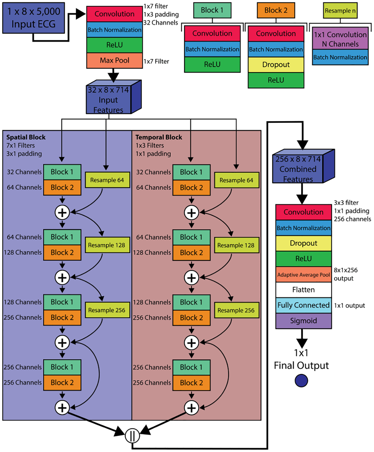
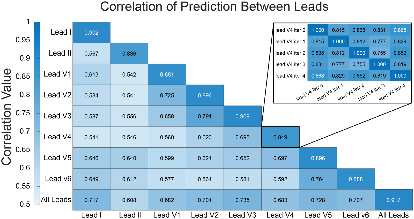
The 12-lead electrocardiogram (ECG) is the most common front-line diagnosis tool for assessing cardiovascular health, yet traditional ECG analysis cannot detect many diseases. Machine learning (ML) techniques have emerged as a powerful set of techniques for producing automated and robust ECG analysis tools that can often predict diseases and conditions not detectable by traditional ECG analysis. Many contemporary ECG-ML studies have focused on utilizing the full 12-lead ECG; however, with the increased availability of single-lead ECG data from wearable devices, there is a clear motivation to explore the development of single-lead ECG-ML techniques. In this study we developed and applied a deep learning architecture for the detection of low left ventricular ejection fraction (LVEF), and compared the performance of this architecture when it was trained with individual leads of the 12-lead ECG to the performance when trained using the entire 12-lead ECG. We observed that single-lead-trained networks performed similarly to the full 12-lead-trained network. We also noted patterns of agreement and disagreement between network low LVEF predictions across the different lead-trained networks.
Figures
Similar articles
-
Assessing and Mitigating Bias in Medical Artificial Intelligence: The Effects of Race and Ethnicity on a Deep Learning Model for ECG Analysis.Circ Arrhythm Electrophysiol. 2020 Mar;13(3):e007988. doi: 10.1161/CIRCEP.119.007988. Epub 2020 Feb 16. Circ Arrhythm Electrophysiol. 2020. PMID: 32064914 Free PMC article.
-
Machine learning models of 6-lead ECGs for the interpretation of left ventricular hypertrophy (LVH).J Electrocardiol. 2023 Mar-Apr;77:62-67. doi: 10.1016/j.jelectrocard.2022.12.001. Epub 2022 Dec 13. J Electrocardiol. 2023. PMID: 36641988
-
Point-of-care screening for heart failure with reduced ejection fraction using artificial intelligence during ECG-enabled stethoscope examination in London, UK: a prospective, observational, multicentre study.Lancet Digit Health. 2022 Feb;4(2):e117-e125. doi: 10.1016/S2589-7500(21)00256-9. Epub 2022 Jan 5. Lancet Digit Health. 2022. PMID: 34998740 Free PMC article.
-
Searching for the Best Machine Learning Algorithm for the Detection of Left Ventricular Hypertrophy from the ECG: A Review.Bioengineering (Basel). 2024 May 15;11(5):489. doi: 10.3390/bioengineering11050489. Bioengineering (Basel). 2024. PMID: 38790356 Free PMC article. Review.
-
Application of Neural Networks to 12-Lead Electrocardiography - Current Status and Future Directions.Circ Rep. 2019 Nov 2;1(11):481-486. doi: 10.1253/circrep.CR-19-0096. Circ Rep. 2019. PMID: 33693089 Free PMC article. Review.
Cited by
-
A noninvasive hyperkalemia monitoring system for dialysis patients based on a 1D-CNN model and single-lead ECG from wearable devices.Sci Rep. 2025 Jan 23;15(1):2950. doi: 10.1038/s41598-025-85722-8. Sci Rep. 2025. PMID: 39848991 Free PMC article.
References
-
- Rafie N, Kashou AH, Noseworthy PA. Ecg interpretation: Clinical relevance, challenges, and advances. Hearts 2021; 2(4):505–513. ISSN 2673–3846.
-
- Jentzer JC, Kashou AH, Attia ZI, Lopez-Jimenez F, Kapa S, Friedman PA, Noseworthy PA. Left ventricular systolic dysfunction identification using artificial intelligence-augmented electrocardiogram in cardiac intensive care unit patients. International jJournal of Cardiology 3 2021; 326:114–123. ISSN 1874–1754. - PubMed
-
- Mahayni AA, Attia ZI, Medina-Inojosa JR, Elsisy MF, Noseworthy PA, Lopez-Jimenez F, Kapa S, Asirvatham SJ, Friedman PA, Crestenallo JA, Alkhouli M. Electrocardiography-based artificial intelligence algorithm aids in prediction of long-term mortality after cardiac surgery. Mayo Clinic Proceedings 12 2021;96:3062–3070. ISSN 1942–5546. - PubMed
Grants and funding
LinkOut - more resources
Full Text Sources