. 2024 Aug 23:19:8661-8679.
doi: 10.2147/IJN.S469687. eCollection 2024.

In vitro/In vivo Evaluations of Hydroxyapatite Nanoparticles with Different Geometry

Affiliations

In vitro/In vivo Evaluations of Hydroxyapatite Nanoparticles with Different Geometry

Weitang Sun et al. Int J Nanomedicine. .

Abstract

Purpose: Hydroxyapatite-based nanoparticles have found diverse applications in drug delivery, gene carriers, diagnostics, bioimaging and tissue engineering, owing to their ability to easily enter the bloodstream and target specific sites. However, there is limited understanding of the potential adverse effects and molecular mechanisms of these nanoparticles with varying geometries upon their entry into the bloodstream. Here, we used two commercially available hydroxyapatite nanoparticles (HANPs) with different geometries (less than 100 nm in size each) to investigate this issue.

Methods: First, the particle size, Zeta potential, and surface morphology of nano-hydroxyapatite were characterized. Subsequently, the effects of 2~2000 μM nano-hydroxyapatite on the proliferation, migration, cell cycle distribution, and apoptosis levels of umbilical vein endothelial cells were evaluated. Additionally, the impact of nanoparticles of various shapes on the differential expression of genes was investigated using transcriptome sequencing. Additionally, we investigated the in vivo biocompatibility of HANPs through gavage administration of nanohydroxyapatite in mice.

Results: Our results demonstrate that while rod-shaped HANPs promote proliferation in Human Umbilical Vein Endothelial Cell (HUVEC) monolayers at 200 μM, sphere-shaped HANPs exhibit significant toxicity to these monolayers at the same concentration, inducing apoptosis/necrosis and S-phase cell cycle arrest through inflammation. Additionally, sphere-shaped HANPs enhance SULT1A3 levels relative to rod-shaped HANPs, facilitating chemical carcinogenesis-DNA adduct signaling pathways in HUVEC monolayers. In vivo experiments have shown that while HANPs can influence the number of blood cells and comprehensive metabolic indicators in blood, they do not exhibit significant toxicity.

Conclusion: In conclusion, this study has demonstrated that the geometry and surface area of HANPs significantly affect VEC survival status and proliferation. These findings hold significant implications for the optimization of biomaterials in cell engineering applications.

Keywords: biocompatibility; circulatory system; hydroxyapatite nanoparticle; proliferation; survival.


Conflict of interest statement

The authors report no conflicts of interest in this work.

Figures

Figure 1
Figure 1
Characterization of rods and spheres HAPNs. (A) Representative TEM images of rods and spheres HAPNs with different magnifications; the inset in right panel showed the corresponding SAED pattern of rods and spheres HAPNs. (B) The number-average size distribution of rods and spheres HAPNs. (C) XRD pattern of rods and spheres HAPNs powder. (D) XPS spectra of the rods and spheres HAPNs Figure powder. (E) FTIR spectra of the rods and spheres HAPNs powder. (F) Raman spectra of freshly prepared rods and spheres HAPNs after storage in ddH2O for 2 days.
Figure 2
Figure 2
Cytotoxicity and mechanisms of HUVEC monolayer following exposure to rods or spheres HAPNs. (A) Cell viability of HUVEC monolayers treated with rods and spheres HAPNs with various concentrations after 24 h. Data are presented as mean ± S.D. (n = 3). (B) Representative fluorescence micrographs of HUVEC monolayers stained with calcein AM (live cells, green fluorescence) and PI (dead cells, red fluorescence) after various treatments, scale bars, 20 μm. Experiments were treated three times. (C) Representative flow cytometer results of AnnexinV-FITC/PI double-stained cells after various treatments. (D) Histogram depicting of the cell population distribution in (C). (E) Representative cell cycle analysis of cells after various treatments. (F) Histogram depicting of the cell population distribution in cell cycle phase in (E). Data are presented as mean ± S.D. (n = 3). Statistical differences were calculated via one-way ANOV A with a Tukey post-hoc test for multiple comparisons. *p < 0.05, **p < 0.01, ***p < 0.001.
Figure 3
Figure 3
Proliferative function of HUVEC monolayers after exposure to rods or spheres HAPNs. A, B, Representative immunostaining of Ki67+ (A) and relative quantification of Ki67+ (B) in HUVEC monolayers after various treatments. (C and D) Representative transwell analysis images (C) and relative quantification of migrated HUVECs from HUVEC monolayer (D) after various treatments. E, F, Representative micrographs of HUVEC monolayer scratch at 0 h (upper panels) and 24 h (lower panels) (E) and relative quantification of cells (F) that migrated back into the wound after various treatments. Scale bar, (A) 20 μm; (C) 200 μm; (E) 400 μm. Data are presented as mean ± S.D. (n = 3). Statistical differences were calculated via one-way ANOVA with a Tukey post-hoc test for multiple comparisons. *p < 0.05, **p < 0.01, ***p < 0.001.
Figure 4
Figure 4
Gene expression pattern of HUVEC monolayer following exposure to rods or spheres HAPNs. (A) Correlation heat map of the tested samples. (B) PCA analysis of the tested samples. (C) Volcano plot of the DECs after two groups comparison. (D) Heat map of DECs of the tested samples. (E) Correlation heat map of DEGs.
Figure 5
Figure 5
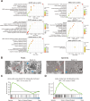
Enrichment analysis and validation of HUVEC monolayer following exposure to rods or spheres HAPNs. (A) Top 10 enriched GO biological process (BP) terms and KEGG pathway of DEGs after two groups comparison. (B) Representative TEM micrographs of HAPN treated HUVEC monolayer. Left panels are the whole-cell micrographs of rods and spheres HAPNs treated HUVEC monolayers, right panels are enlarged micrographs of red dotted line in left panels. Blue arrows indicate the mitochondria, yellow arrow indicates nuclear cleavage. Scale bars: left panels, 5 μm; right panels, 500 nm. (C and D) GSEA analysis of NOD-like receptor signaling pathway (C) and oxidative phosphorylation (D) in spheres HAPN-treated HUVEC monolayers compared with rods HAPN-treated HUVEC monolayers.
Figure 6
Figure 6
In vivo cardiovascular safety assessment after exposure to rods or spheres HAPNs. A, Representative ECGs of mice at 1, 7, and 14 days after gavage of saline, rods and spheres HAPNs. B, C, Heat map of representative haematological data (B) and serum biochemistry data (C) of mice with rods or spheres HAPNs at 14 days post-gavage. Data are displayed as relative quantification (RQ) to saline-treated mice. D, E, H&E staining of the heart, liver, kidney, spleen and lung at low magnification (D) and high magnification (E) of the saline, rods, or spheres HAPNs at 21 days post-gavage. Scar bars: (D),1000 μm; (E) 100 μm. The black box in (D) indicates the area of enlarged images in (E).
Figure 7
Figure 7
Sphere-shaped hydroxyapatite nanoparticles with a higher surface area induce inflammatory apoptosis/necrosis in vascular endothelial cells monolayer.

References

    1. Qi C, Musetti S, Fu LH, Zhu YJ, Huang L. Biomolecule-assisted green synthesis of nanostructured calcium phosphates and their biomedical applications. Chem Soc Rev. 2019;48(10):2698–2737. doi:10.1039/c8cs00489g - DOI - PubMed
    1. Ethirajan A, Ziener U, Chuvilin A, Kaiser U, Cölfen H, Landfester K. Biomimetic Hydroxyapatite Crystallization in Gelatin Nanoparticles Synthesized Using a Miniemulsion Process. Adv Funct Mater. 2008;18(15):2221–2227. doi:10.1002/adfm.200800048 - DOI
    1. Bera B, Saha Chowdhury S, Sonawane VR, De S. High capacity aluminium substituted hydroxyapatite incorporated granular wood charcoal (Al-HApC) for fluoride removal from aqueous medium: batch and column study. Chem Eng J. 2023;466. doi:10.1016/j.cej.2023.143264 - DOI
    1. Zhao H, Wu C, Gao D, et al. Antitumor Effect by Hydroxyapatite Nanospheres: activation of Mitochondria-Dependent Apoptosis and Negative Regulation of Phosphatidylinositol-3-Kinase/Protein Kinase B Pathway. ACS nano. 2018;12(8):7838–7854. doi:10.1021/acsnano.8b01996 - DOI - PubMed
    1. Ding ZZ, Fan ZH, Huang XW, et al. Bioactive Natural Protein-Hydroxyapatite Nanocarriers for Optimizing Osteogenic Differentiation of Mesenchymal Stem Cells. J Mat Chem B. 2016;4(20):3555–3561. doi: 10.1039/c6tb00509h - DOI - PMC - PubMed

LinkOut - more resources