. 2024 Dec;13(1):2396870.
doi: 10.1080/22221751.2024.2396870. Epub 2024 Sep 5.

Genomic characterization of an emerging Rickettsia barbariae isolated from tick eggs in northwestern China

Affiliations

Genomic characterization of an emerging Rickettsia barbariae isolated from tick eggs in northwestern China

Ning Wang et al. Emerg Microbes Infect. 2024 Dec.

Abstract

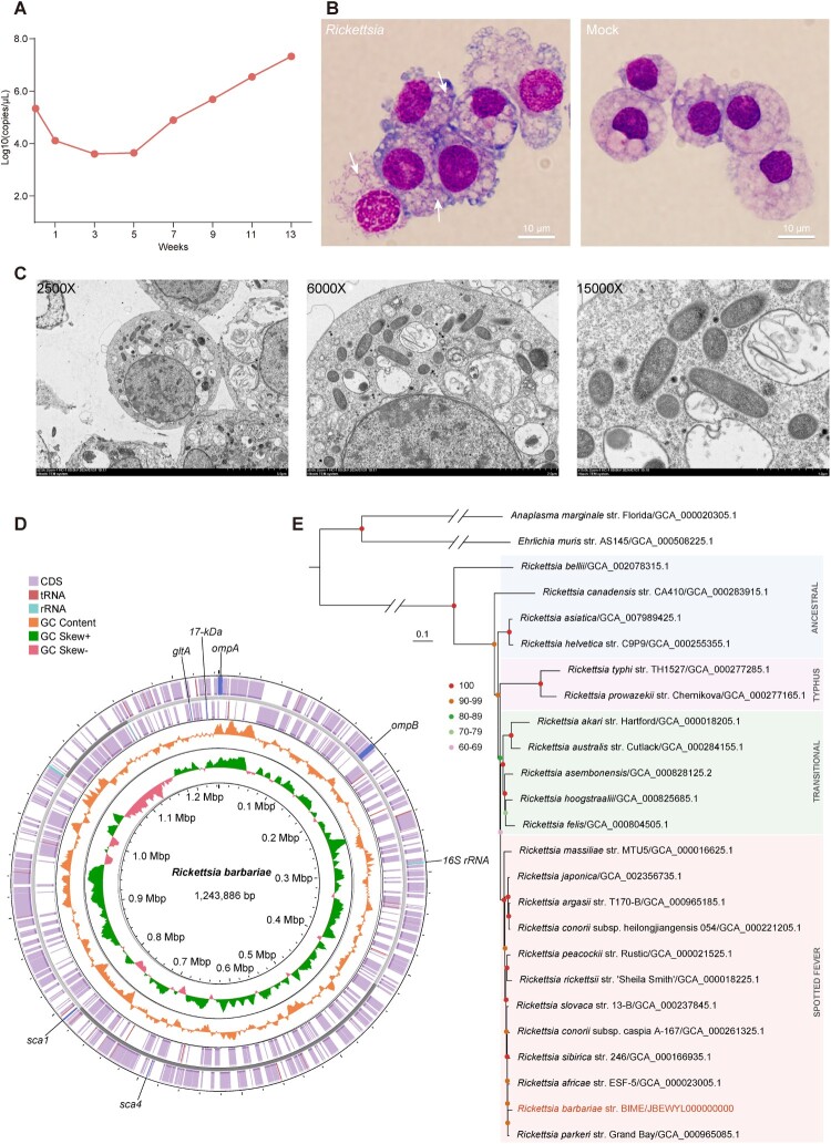
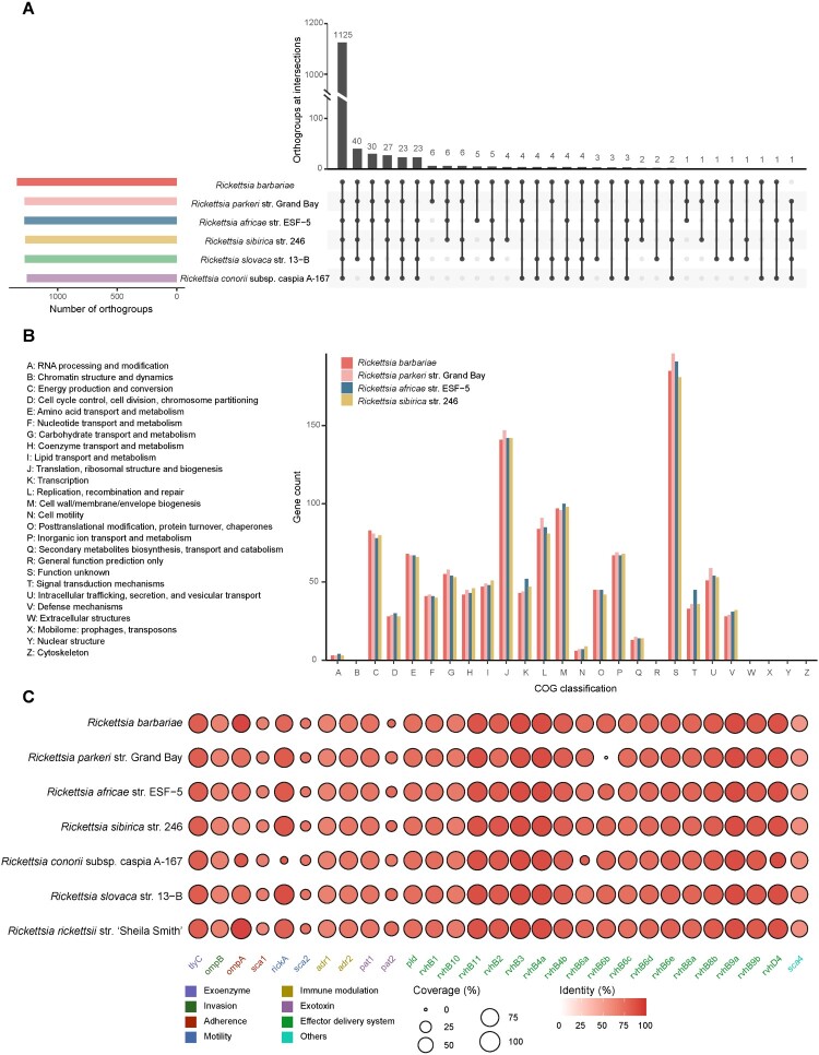
The continual emergence of tick-borne rickettsioses has garnered widespread global attention. Candidatus Rickettsia barbariae (Candidatus R. barbariae), which emerged in Italy in 2008, has been detected in humans from northwestern China. However, the lack of Candidatus R. barbariae genome and isolated strains limits the understanding of its biological characteristics and genomic features. Here, we isolated the Rickettsia for the first time from eggs of Rhipicephalus turanicus in northwestern China, and assembled its whole genome after next-generation sequencing, so we modified the proposed name to Rickettsia barbariae (R. barbariae) to conform to the International Code of Nomenclature of Prokaryotes. Phylogenetic analysis based on the whole genome revealed that it was most closely related to the pathogenic Rickettsia parkeri and Rickettsia africae. All virulence factors, present in the pathogenic spotted fever group rickettsiae, were identified in the R. barbariae isolate. These findings highlight the pathogenic potential of R. barbariae and the necessity for enhanced surveillance of the emerging Rickettsia in the human population.

Keywords: China; Rhipicephalus turanicus; Rickettsia barbariae; phylogenetic analysis; virulence factors; whole-genome sequencing.


Conflict of interest statement

No potential conflict of interest was reported by the author(s).

Figures

Figure 1.
Figure 1.
Isolation and fundamental genomic information of R. barbariae str. BIME. (A) Growth curves of R. barbariae in IDE8 tick cells over 13 weeks after inoculated the eggs of Rh. turanicus onto IDE8 cells. (B) Giemsa staining of R. barbariae isolated from the eggs of Rh. turanicus in IDE8 cells. Rickettsia appeared as deeply stained, short-rod, long-rod, or spherical bodies in shades of blue-purple. The uninfected IDE8 cells cultivated in parallel were used as controls. Scale bar represents 10 μm. (C) Transmission electron micrographs of IDE8 cells infected with R. barbariae. Photomicrographs were captured with an HT7800 transmission electron microscope camera. Scale bar represents 5 μm (magnification, × 2,500), 2 μm (magnification, × 6,000) and 1 μm (magnification, × 15,000). (D) Circular map of R. barbariae str. BIME genome. From inner circle to outer circle representative GC skew, GC content, proteins of – strand, contig, proteins of + strand. The location of tRNA, rRNA, ompA, ompB, gltA, 17-kDa, sca1, sca4 and 16S rRNA genes within the genome are indicated. (E) Phylogenetic tree constructed using the maximum likelihood method with 1,000 replications, based on the whole genomes of 24 other publicly available established or proposed Rickettsiales species. Anaplasma marginale and Ehrlichia muris were used as outgroup species. Scale bar indicates 0.1 nucleotide substitutions per site.
Figure 2.
Figure 2.
Functional annotation of R. barbariae str. BIME genome. (A) UpsetR plot showing the number of orthogroups in the genome of R. barbariae compared with other closely related SFGR representatives. Connected circles indicate shared orthogroups among these SFGR species. (B) COG annotation of R. barbariae and other closely related SFGR genomes. (C) Heatmap illustrating the presence of virulence factors in R. barbariae and other representative SFGR genomes. The vertical axis represents various SFGRs, the horizontal axis indicates the names of the virulence factors, the size of the bubbles represents the coverage of the virulence factors, and the colour intensity indicates their identity.
Figure 3.
Figure 3.
Phylogenetic analysis of Rickettsia barbariae based on the nucleotide sequences of five genes. (A) Phylogenetic tree based on the ompA gene. (B) Phylogenetic tree based on the ompB gene. (C) Phylogenetic tree based on the gltA gene. (D) Phylogenetic tree based on the sca1 gene. (E) Phylogenetic tree based on the sca4 gene. Bootstrap analysis with 1,000 replicates was conducted to evaluate phylogenetic robustness. Scale bar indicates the number of nucleotide substitutions per site. Sequences highlighted in red font represent those obtained in this study, while those in blue font denote Candidatus R. barbariae sequences identified from human samples in GenBank.

Similar articles

Cited by

References

    1. Seidi S, Omidi AH, Esmaeili S.. Distribution of different Rickettsia species in countries of the WHO Eastern Mediterranean (WHO-EMRO) region: An overview. Travel Med Infect Dis. 2024;58:102695. doi:10.1016/j.tmaid.2024.102695 - DOI - PubMed
    1. Fang LQ, Liu K, Li XL, et al. . Emerging tick-borne infections in mainland China: an increasing public health threat. Lancet Infect Dis. 2015;15:1467–1479. doi:10.1016/s1473-3099(15)00177-2 - DOI - PMC - PubMed
    1. Zhang YY, Sun YQ, Chen JJ, et al. . Mapping the global distribution of spotted fever group rickettsiae: a systematic review with modelling analysis. Lancet Digit Health. 2023;5:e5–e15. doi:10.1016/s2589-7500(22)00212-6 - DOI - PMC - PubMed
    1. Mura A, Masala G, Tola S, et al. . First direct detection of rickettsial pathogens and a new rickettsia, ‘Candidatus rickettsia barbariae’, in ticks from Sardinia, Italy. Clin Microbiol Infect. 2008;14:1028–1033. doi:10.1111/j.1469-0691.2008.02082.x - DOI - PubMed
    1. Cicculli V, Capai L, Quilichini Y, et al. . Molecular investigation of tick-borne pathogens in ixodid ticks infesting domestic animals (cattle and sheep) and small rodents (black rats) of Corsica, France. Ticks Tick Borne Dis. 2019;10:606–613. doi:10.1016/j.ttbdis.2019.02.007 - DOI - PubMed

Substances

Supplementary concepts

LinkOut - more resources