Review
. 2024 Aug 28:15910199241272718.
doi: 10.1177/15910199241272718. Online ahead of print.

Rare neurovascular variants that you probably have not seen before

Affiliations
Review

Rare neurovascular variants that you probably have not seen before

Mohamad Abdalkader et al. Interv Neuroradiol. .

Abstract

Background: Recognition of neurovascular variants is crucial for safe endovascular and neurosurgical interventions. We aim to review and highlight various uncommon neurovascular variants and anomalies with a discussion of their relevant embryology and pathology.

Methods: A retrospective review of a prospectively maintained neurovascular database was performed to identify uncommon neurovascular variants and anomalies. A pictorial review of these neurovascular findings is provided along with relevant embryological development, clinical significance, and potential pathological associations.

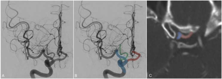
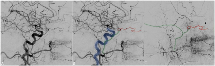
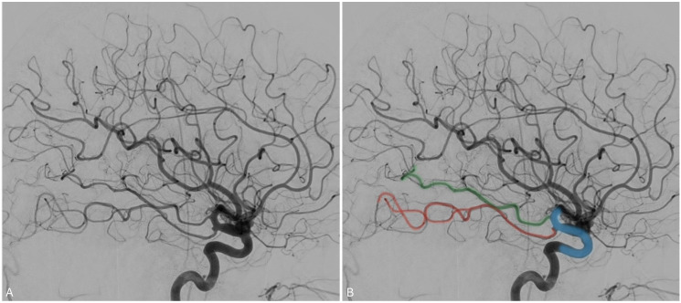
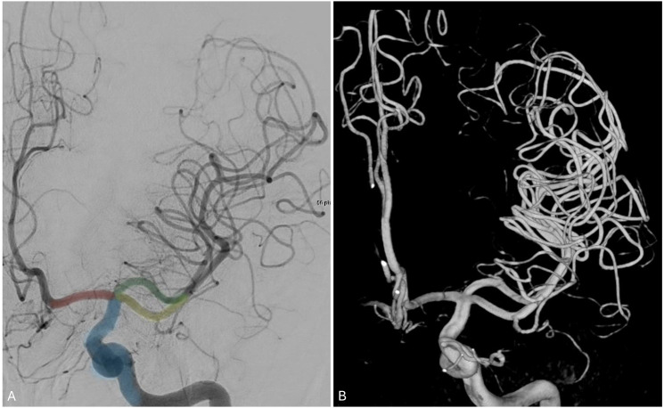
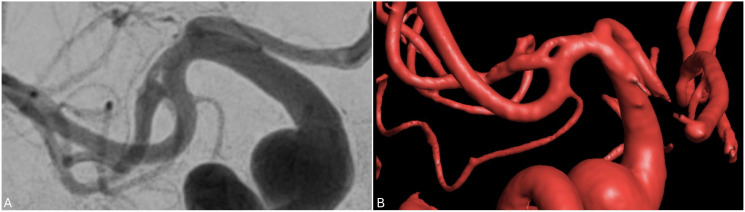
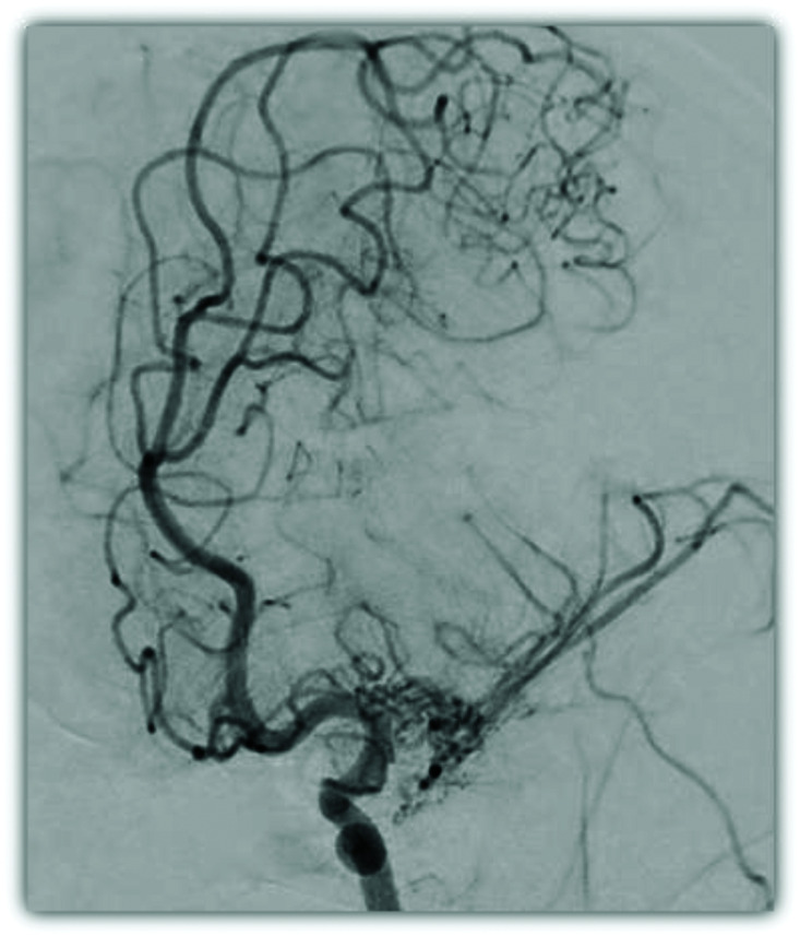
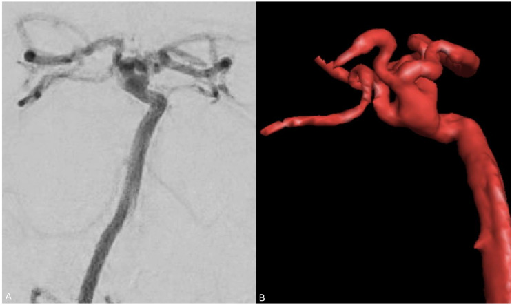
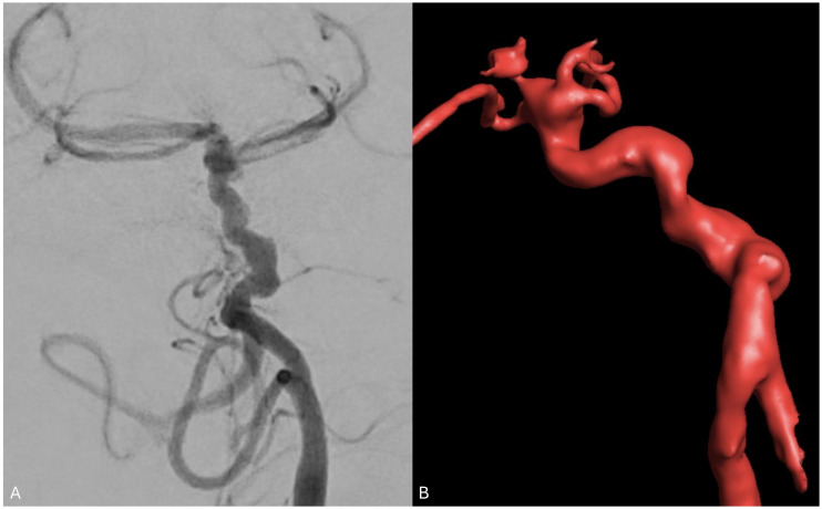
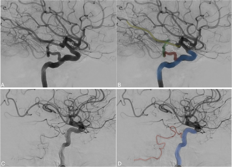
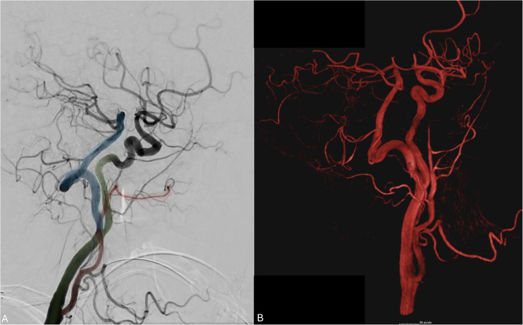
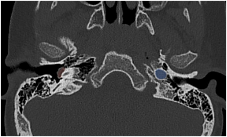
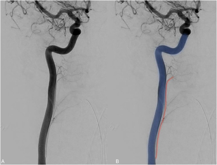
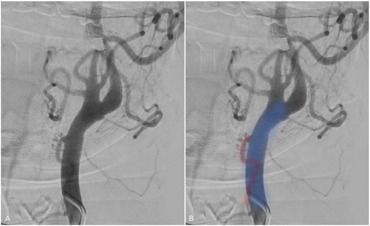
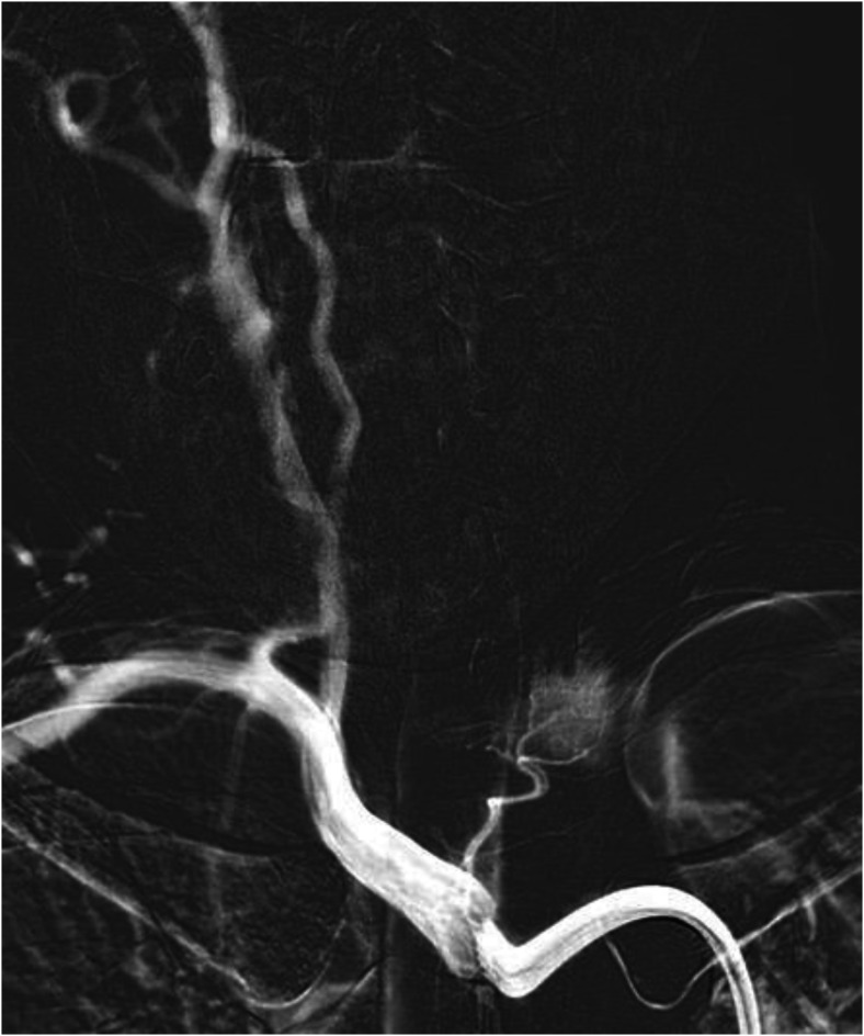
Results: A pictorial review of selected neurovascular variants and anomalies is presented. These entities, divided between intra- and extra-cranial findings, include infra-optic origin of the anterior cerebral artery, meningo-ophtalmic artery, duplicated posterior cerebral artery, duplicate middle cerebral artery (MCA), MCA fenestration, twig-like MCA, pure arterial malformation, corkscrew basilar artery, persistent hypoglossal artery, persistent trigeminal artery and its variants, direct branches from the common carotid and cervical internal carotid arteries (ICA) (ascending pharyngeal artery from the ICA, thyroidal arteries from the CCA/brachiocephalic, arteria thyroidea ima), and extra-cranial carotid fenestration. The angiographic findings of these entities are presented with relevant 3D reconstruction and multimodal cross-sectional imaging correlation when available.

Conclusions: This pictorial review highlights uncommon neurovascular variants and anomalies that neuroradiologists, interventionalists, and neurosurgeons should be aware of for accurate diagnosis and safe interventions.

Keywords: Neurovascular variants; angiography; arterial variants; neurointervention; variant anatomy.


Conflict of interest statement

Declaration of conflicting interestsThe authors declared no potential conflicts of interest with respect to the research, authorship, and/or publication of this article.

Figures

Figure 1.
Figure 1.
Angiogram of the right internal carotid artery and axial CT angiogram demonstrating an infra-optic anterior cerebral artery (red) arising from the anterior genu of the ICA (blue). A posterior communicating artery (blue) is noted arising from its expected location of the supraclinoid ICA.
Figure 2.
Figure 2.
A. Angiogram of the right common carotid artery demonstrating no ophthalmic artery from the ICA (blue). The OphA (red) originates from the MMA (green). This is better seen in the selective angiogram of the external carotid artery (C). The choroidal artery blush (black arrow on C) is noted from the selective external carotid artery injection.
Figure 3.
Figure 3.
Angiogram of the right ICA in the lateral projection demonstrating a true fetal PCA, or “duplicated PCA” (red and green).
Figure 4.
Figure 4.
(a) angiogram in the anteroposterior projection showing the ICA (blue) giving rise to the left ACA (red) and the two left MCA branches (superior division in green and inferior division in yellow). (b) A 3D reconstruction of the angiogram.
Figure 5.
Figure 5.
Anteroposterior and 3D reconstruction of an angiogram showing a right MCA fenestration.
Figure 6.
Figure 6.
Angiogram in oblique orientation demonstrating the plexiform vessels constituting a left T-MCA with delay filling of the MCA territory compared to the ACA territory.
Figure 7.
Figure 7.
Angiogram of the vertebral artery with 3D volume rendering reconstruction demonstrating a PAM of the distal basilar artery with coiled and tortuous basilar tip involving the PCA and SCA branches.
Figure 8.
Figure 8.
Angiogram of the vertebral artery with 3D volume rendering reconstruction demonstrating tortuosity with many distal angular turns of the basilar artery consisting with CBA. This patient presented with multiple intracranial anterior circulation aneurysms.
Figure 9.
Figure 9.
(a) lateral angiogram of the ICA (blue) showing a PTA (red) anastomosing the ICA to the basilar artery. Coil-embolized aneurysms can be seen in the posterior and anterior communicating arteries. (b) Lateral angiogram of the ICA (blue) showing a PTA variant contributing to the anterior inferior cerebellar territory as cerebellar artery (red).
Figure 10.
Figure 10.
Lateral common carotid angiogram and 3D angiogram demonstrating a PHA (blue) persistent anastomosis between the ICA (green) and the BA. The external carotid artery is noted in red.
Figure 11.
Figure 11.
An axial CT scan showing a right-sided aberrant ICA (highlighted in red) protruding in the tympanic fossa. The left ICA is highlighted in blue for comparison.
Figure 12.
Figure 12.
Right ICA angiogram demonstrating the pharyngeal branch of the ascending pharyngeal artery (red) branching off the ICA (blue).
Figure 13.
Figure 13.
Angiogram demonstrating superior thyroidal artery (red) arising from the CCA (blue) proximal to the carotid bifurcation.
Figure 14.
Figure 14.
Angiogram of the brachiocephalic artery demonstrating a thyroid ima artery arising from the proximal brachiocephalic trunk.

References

    1. Bonasia S, Smajda S, Ciccio G, et al. Embryology of the anterior communicating artery complex: implications on possible adult variants. Surg Radiol Anat SRA 2022; 44: 737–748. - PubMed
    1. Nutik S, Dilenge D. Carotid-anterior cerebral artery anastomosis. Case report. J Neurosurg 1976; 44: 378–382. - PubMed
    1. Tahir RA, Haider S, Kole M, et al. Anterior cerebral artery: variant anatomy and pathology. J Vasc Interv Neurol 2019; 10: 16–22. - PMC - PubMed
    1. Shapiro M, Sharashidze V, Nossek E, et al. Superior hypophyseal arteries: angiographic re-discovery, comprehensive assessment, and embryologic implications. J NeuroInterventional Surg 2023; jnis-2023-020922. - PubMed
    1. Toma N. Anatomy of the ophthalmic artery: embryological consideration. Neurol Med Chir (Tokyo) 2016; 56: 585–591. - PMC - PubMed

LinkOut - more resources